A 31-year-old previously healthy man comes to your office because of a 2-week history of low-grade fevers, weight loss, malaise, nocturnal tightness in his chest, and shortness of breath. He also reports a small amount of leg swelling and scrotal swelling over this time. Prior to 2 weeks ago, he denies any recent illnesses, sick contacts, or travel. He also denies any hemoptysis or sinus infections. His temperature is 38.0 C (100.2 F), blood pressure is 170/95 mm Hg, pulse is 77/min, and respirations are 14/min. Physical examination shows trace bilateral lower extremity and scrotal edema. Bilateral wheezes are also appreciated. A chest x-ray is unremarkable. Laboratory studies show:

Sodium141 mEq/dL
Potassium5.1 mEq/dL
Chloride98 mEq/dL
Bicarbonate21 mEq/dL
Urea nitrogen, serum21 mg/dL
Creatinine2.5 mg/dL
Glucose97 mg/dL
Serum eosinophilsmildly elevated

Urinalysis
Colorclear
Specific gravity1.020
Osmolality55 mOsmol/kg
Leukocyte esterasenegative
Nitritenegative
Protein2+
Blood4+
Microscopicmany red cell casts.
24 hour urine protein collection1900 mg

The laboratory finding that would support the most likely diagnosis is

  A. anti-glomerular basement membrane antibody
  B. antistreptolysin titer (ASO)
  C. cANCA
  D. elevated serum IgA levels
  E. pANCA
Explanation:

The correct answer is E. This patient likely has Chrug-Struass syndrome. Chrug-Struass syndrome is a vasculitis, which is associated with eosinophilia and asthma. This patient has nephritic syndrome as evidenced by edema, hypertension, and hematuria. Asthma is suggested by his nocturnal chest tightness and shortness of breath. Laboratory results reveal elevated eosinophils, which is typical in this condition. Chrug-Strauss is associated with a positive pANCA.

Goodpastures syndrome is typically defined as a combination of nephritic syndrome and pulmonary hemorrhage, although some patients have nephritic syndrome alone. The disease is mediated by antibodies to the glomerular basement membrane (choice A) and therefore those are the laboratory results often associated with the disease.

ASO (choice B) is associated with post infectious glomerulonephritis. Patients present with oliguria, hypertension, and edema. They classically complain of Coca-Cola colored urine. These infections classically occur 1-3 weeks after infection with nephritogenic group A streptococci (step throat or cellulitis).

Wegner granulomatosis is similar to Chrug-Strauss but respiratory involvement such as nasal septal perforation or sinus problems are typical presenting signs. Wegners doesn't have an association with asthma or increased serum eosinophils. The test associated with Wegners is cANCA (choice C).

Bergers syndrome (IgA nephropathy) presents in a similar manner to post-infectious glomerulonephritis but there is no latent period between infection and kidney involvement. These patients typically have gross hematuria after a viral illness. About 50% of these patients will have an elevated IgA (choice D).


A 22-year-old medical student has a PPD placed at his yearly physical exam. He has no medical problems and is currently taking no medications. He returns to your office three days after his initial appointment with 8 mm of induration around his PPD. He is concerned that he is infected with tuberculosis. At this time you should

  A. begin sputum collection for AFB staining and culture
  B. explain that he needs an extended course of single drug therapy
  C. explain that he needs an extended course of four drug therapy
  D. explain that no intervention is indicated at this time
  E. recommend a chest x-ray to determine appropriate treatment
Explanation:

The correct answer is D. When evaluating a patient for a positive tuberculin skin you must first classify which risk category the patient belongs. Patients are divided into three groups: High risk patients are considered to have a positive skin test if the area of induration is greater than or equal to 5 mm. These are people with HIV or those at high risk for HIV (i.e. IV drug users), people who have close contact with patients with active TB, or those who have had TB or evidence of prior TB on chest x-ray. Patients with induration greater than or equal to 10 mm are considered to have a positive test if they immigrated from countries with a high prevalence of TB, are known HIV negative IV drugs users, are medically underserved populations, prisoners, mental institution residents, nursing home residents, those with some chronic medical problems, or hospital workers. All other people are considered to have a positive test if the area of induration is greater than 15 mm. Our patient has an area of induration of less than 10 mm. No further work up is needed in this patient.

Collecting sputum for acid fast bacilli (AFB) (choice A) is helpful in confirming the diagnosis of suspected tuberculosis. If this patient reported typical symptoms of tuberculosis such as fevers, night sweats, weight loss, and cough a positive AFB smear or culture would help us to make the diagnosis of TB.

Extended course of single drug therapy (choice B) would be necessary if this patient had a positive skin test with no evidence of active infection. Preventative therapy has been shown to reduce the incidence of reactivated tuberculosis.

An extended course of four-drug therapy (choice C) is the treatment of choice for active tuberculosis. A typical four-drug regimen includes isoniazid, rifampin, pyrazinamide, and ethambutol or streptomycin.

A recent converter of their skin test should get a chest x-ray (choice E) to evaluate for any evidence of active disease and to have a baseline chest x-ray that could be followed in the future. Once a patient has a positive skin test, it is no longer valuable to perform further skin tests. Rather, yearly chest x-rays for evidence of reactivated TB should be done.


A 26-year-old woman comes to the office for routine postpartum care after delivering a healthy baby boy 6 days earlier. This is her first child and she is concerned about her breastfeeding, diaper changing, and bathing techniques. You ask her to explain how she performs these activities, and it seems as if she is doing everything correctly. She then tells you that she is very upset about her weight, that she gained 45 pounds during the pregnancy, and now is still 37 pounds "overweight". Her husband is a good friend of yours from medical school. Her blood pressure is 140/90 mm Hg and her pulse is 95/min. Physical examination shows dilated pupils, but is otherwise unremarkable. You suspect that she is using drugs and so you ask her in a direct, nonjudgmental way. She admits to using cocaine twice since the delivery of her son. The most appropriate next step is to

  A. advise her to stay away from her son when she is using cocaine
  B. call her husband and ask if he knows that she is using drugs
  C. contact the child welfare association and inform them of your findings
  D. obtain a urine sample to test for cocaine and other illegal drugs
  E. tell her that she should not breast feed her son if she is using cocaine
Explanation:

The correct answer is E. Cocaine is contraindicated during breast feeding and so you need to advise the mother to stop breast feeding if she is using cocaine. Possible effects of cocaine in the breast milk include growth retardation and neurologic damage. Cocaine use during pregnancy is associated with fetal demise, spontaneous abortions, premature rupture of membranes, preterm labor and delivery, placental abruption, intrauterine growth retardation, and congenital anomalies. Other medications and drugs that are contraindicated during breastfeeding are bromocriptine, cyclophosphamide, cyclosporine, doxorubicin, lithium, ergotamine, methotrexate, amphetamines, heroin, marijuana, nicotine, and phencyclidine.

The most appropriate next step is to tell her is to discontinue breastfeeding if she is using cocaine, not to advise her to stay away from her son when she is using cocaine (choice A). This answer makes it seem like it is okay that she is using drugs, but she should just not be near her baby. This is actually not true. If she is using the cocaine to lose weight, you should recommend a healthy diet and exercise regimen.

It is incorrect to call her husband and ask if he knows that she is using drugs (choice B) or to contact the child welfare association and inform them of your findings (choice C). You should first try to reason with the patient and encourage her to discontinue using cocaine and tell her that she is not only risking her own life now, but also that of her son's because she is breast feeding.

She already admitted to you that she is using cocaine and so it is unnecessary to obtain a urine sample to test for cocaine and other illegal drugs (choice D).


A 28-year-old woman comes to the office because of a 4-day history of "itching, burning, and an awful-smelling vaginal discharge". She says that she and her boyfriend had similar symptoms a few months ago, which resolved after treatment by his physician. Now, she believes that he is "fooling around," because this "disease" has returned. On physical examination her vulva is erythematous and there are patches of petechiae in the upper vagina and on the cervix. There is a copious amount of yellowish-green, "frothy", malodorous vaginal discharge. Examination of the discharge on a saline wet mount will most likely reveal

  A. branching hyphae and spores
  B. epithelial cells with clumps of bacteria and "ground-glass" cytoplasm
  C. giant multinucleated cells with intranuclear inclusions
  D. koilocytes
  E. motile, flagellated organisms
Explanation:

The correct answer is E. This patient has the signs and symptoms most consistent with a Trichomonas vaginalis infection, which is diagnosed by finding motile, flagellated organisms on a saline wet mount smear preparation. Patients with T. vaginalis typically experience vulvar itching and burning, a "frothy" malodorous discharge, dysuria, dyspareunia, and frequency and urgency of urination. Vaginal and cervical petechiae ("strawberry cervix") may be present. The treatment is metronidazole and it is important to treat the partner because T. vaginalis can be transmitted by sex.

Branching hyphae and spores (choice A) are associated with an infection with Candida albicans, which is characterized by intense pruritus and a thick, white ("cottage-cheese") discharge. This patient's discharge is not consistent with a Candida infection.

Epithelial cells with clumps of bacteria and "ground-glass" cytoplasm (choice B) are associated with an infection with Gardnerella vaginalis, which is characterized by gray-white, "fishy" discharge. Irritation of the vaginal epithelium is not usually seen. This patient's discharge is not consistent with a Gardnerella infection.

Giant multinucleated cells with intranuclear inclusions (choice C) are associated with an infection with herpes simplex virus, which is characterized by vesicular lesions, ulcers, paraesthesia, and dysuria. The diagnosis is confirmed with viral cultures and scrapings. Giant multinucleated cells with eosinophilic intranuclear inclusions are seen when stained with Wright's stain. A saline wet mount smear preparation is not used to diagnose herpes infections. Also, this patient's discharge is not consistent with a herpes infection.

Koilocytes (choice D) are associated with an infection with the human papilloma virus (HPV) which is characterized by soft, fleshy lesions on the genital region (condyloma acuminata). The diagnosis is established with a biopsy of the lesions. A Pap smear may show "koilocytes", which are cytologic changes associated with HPV. A saline wet mount smear preparation is not used to diagnose HPV. This patient's signs and symptoms are inconsistent with HPV.


A 20-year-old Hispanic man recently discovered changes in his skin that he describes as "ugly spots with terrible itching". He has a history of a short-lived substance abuse problem when he was 18 years old, which he got help for immediately. Although he is now "clean" he wonders if this may be the cause of all his skin problems. The patient's father, who presently has a drug and alcohol problem, has the same spots on his skin which come and go. On physical examination, there are 2 scaly plaques on the left temporal scalp, his ears are scaly throughout the external auditory canals bilaterally, there are pink, scaly well-defined plaques on his elbows, and distal onycholysis. The next best step to take in an effort to diagnose this condition is to

  A. biopsy one of the marks of the nails
  B. biopsy one of the scalp lesions
  C. biopsy one of the tongue lesions
  D. cut a nail with onycholysis and perform a fungal culture
  E. scrape the skin in the ears and examine with KOH (potassium hydroxide)
Explanation:

The correct answer is B. Although this patient is presenting with many of the classic signs of chronic plaque psoriasis, it is best to document it clearly in the chart with a representative biopsy at least once in a patient with this chronic disease. The easiest place to do a biopsy in this patient is either the scalp or the elbow (not a given choice). The results of a nail biopsy in psoriasis is not as classic (choice A) and a tongue biopsy is difficult compared to other easily accessible skin areas,(choice C). The classic signs of chronic plaque psoriasis are silvery or pink well-defined plaques, which can span the whole body from the scalp to the feet. The most classically involved areas include the scalp, ears, elbows, knees, sacrum and ankles. Psoriasis of the tongue is a migratory glossitis, which is a thickened area of the tongue that is transient. One day it will be in one area of the tongue, the next day, it will move, thus "migratory", to another area on the tongue. Psoriasis of the nails consists of distal onycholysis (distal lifting of the nail plate), subungual debris (scale underneath the nail), total nail dystrophy, oil-spots (again, scale under the nail), and pitting. Pitting can be seen in one or 2 other dermatologic conditions but more often represents psoriasis. Psoriasis is an inherited disease whose exact chromosomal location and mechanism is still under debate. Psoriasis has not been directly linked to substance abuse problems. This patient most likely inherited the psoriasis and perhaps even the substance abuse problems from his father, albeit separately.

There are other forms of psoriasis which include erythrodermic psoriasis which is total body erythema and scaling, which can occur as the first onset of psoriasis or as an exacerbation of existing psoriasis, pustular psoriasis which are crops of sterile pustules, which erupt and can form lakes of pus, and inverse psoriasis which is psoriasis of the groin and intertriginous areas. Because the plaques are constantly moist, they do not have the typical silvery, heaped-up scale of psoriasis. They are still very well-defined and may have some scale on the outer edge of each plaque. Guttate psoriasis is an eruption of "rain-drop" like scaly plaques often seen in children after a group A streptococcal infection.

Although tinea of the skin and nails is in the differential in psoriasis, this patient with so many classic psoriasis findings does not need a KOH or fungal culture of the nails (choice D) or skin from the ear (choice E). Diagnostic tests for fungus are only performed if the lesions are not classic, and the clinical picture is hazy between psoriasis and tinea. The major diagnoses that can cause scaling in the ear are psoriasis and discoid lupus, but rarely fungus.


A 55-year-old man with diabetes is referred to you for the evaluation of chest pain and dyspnea. His medications include glyburide and aspirin. He is afebrile, has a blood pressure of 130/78 mm Hg, heart rate of 72/min, and respiratory rate of 18 /min. His lungs are clear bilaterally. He has a prominent, nondisplaced apical impulse and a I/VI, late peaking systolic ejection murmur at the cardiac base. An echocardiogram result (taken two weeks ago) from the referring physician shows a preserved ejection fraction, mild-moderate concentric hypertrophy without systolic wall motion abnormalities, and an aortic valve area of 0.6 cm2. The most likely cause of the patient's chest pain and dyspnea is

  A. congestive heart failure
  B. critical aortic stenosis
  C. hypertensive cardiomyopathy
  D. idiopathic hypertrophic subaortic stenosis
  E. ischemic heart disease
Explanation:

The correct answer is B. Critical aortic stenosis is suggested by the patient's physical exam (late peaking ejection murmur at the base, prominent apical impulse) and clinched by the echocardiogram showing an aortic valve area of 0.6 cm2 (a valve area less than 0.7 cm2 is considered critical).

Congestive failure (choice A) is unlikely given that there is no mention of the classic physical stigmata (S3, elevated jugular venous pressure, crackles on the lung exam).

Hypertensive cardiomyopathy (choice C) is unlikely given that the patient has no history of preexisting hypertension, and is not hypertensive on exam.

Idiopathic hypertrophic subaortic stenosis (choice D) is unlikely given that there is no mention of outflow obstruction on the echocardiogram.

Ischemic heart disease (choice E) is less likely given the absence of any history of coronary disease, minimal coronary disease risk factors, and the absence of wall motion abnormalities or scar on the echocardiogram.


A 39-year-old man comes to the office because his coworkers have been saying that his face "is lopsided" for the past 2 days. He says that he does not spend much time looking in the mirror so he has not really noticed a cosmetic problem, but he did have pain behind his ear a few days ago and his wife has been making fun of him for drooling lately. He complains that his left eye has been drier than usual and he has had to use lubricating drops. He recently returned from a month long camping and hiking trip through the beautiful wooded regions of Connecticut. You have treated him for contact dermatitis and the "flu" in the past, but you have not seen him in a couple of years. The most appropriate way to test for a facial nerve palsy is to

  A. ask him to clench his teeth while you palpate the masseter muscle
  B. ask him to show you his teeth
  C. have him close his eyes and tell you when you are touching his cheek with gauze
  D. have him turn his head to the right against your hand
  E. touch the posterior pharyngeal wall with an applicator stick
Explanation:

The correct answer is B. This patient's symptoms are consistent with a facial nerve palsy that may be caused by Lyme disease (from his camping trip). To test the facial nerve, you should ask the patient to show you his teeth. If the left side of his face is drawn to the right, he most likely has a left-sided facial palsy. Other facial nerve tests include asking him to wrinkle his forehead, puff out his cheeks, and hold his eyes shut as tightly as possible as you try to carefully pry them open.

If this patient had a trigeminal nerve palsy, you should ask him to clench his teeth while you palpate the masseter muscle (choice A). During this maneuver, his jaw would deviate to the same side as the lesion. Also, he would complain of sensory abnormalities on his forehead, cheek, and jaw and paroxysms of pain on his chin, lips, cheeks, and gums.

Sensory abnormalities can be elicited by having him close his eyes and tell you when you are touching his cheek with gauze (choice C). He does not have these sensory symptoms.

If he had a left-sided spinal accessory nerve palsy, you should have him turn his head to the right against your hand (choice D). The dysfunction would manifest with the patient being unable to do this, or weakness on this side as opposed to the other side. He does not complain of weakness in the head, neck, or shoulder.

The gag reflex of the glossopharyngeal nerve is tested by touch the posterior pharyngeal wall with an applicator stick (choice E). Symptoms of glossopharyngeal nerve lesions are sensory dysfunction of the pharynx, loss of taste on the posterior third of the tongue, and a partially dry mouth.


A 32-year-old woman comes to the office "for a prescription of propranolol for stage fright." She tells you that she is professional singer and lately she has been experiencing "butterflies" and palpitations before performances. She has been so worried about having one of these symptoms that she is having trouble sleeping at night. She tells you that a friend of hers has a similar problem and propranolol has "cured her." She has been a patient of yours for the past 10 years and you remember that she has severe asthma, requiring many hospitalizations, the most recent being 2 weeks ago. Her asthma attacks have been increasingly more severe and have been occurring at an increased frequency. She tells you that she is in a rush and all she needs is the prescription. The most appropriate next step is to

  A. administer a pulmonary function test
  B. explain that propranolol is not a good drug for her
  C. give her a referral to a psychiatrist
  D. order a chest x-ray
  E. prescribe propranolol for her to take before her performances
Explanation:

The correct answer is B. This patient most likely has performance anxiety, which is a form of social phobia. The treatment usually involves beta-blockers before a performance to decrease the symptoms. However, a patient with severe asthma should avoid beta-blockers because they can cause bronchoconstriction and precipitate into an asthmatic attack.

A pulmonary function test (choice A) and a chest x-ray (choice D) are not indicated at this time. You already know that she has asthma that has required hospitalizations and the results of these tests are unlikely to change your management.

A referral to a psychiatrist (choice C) may be helpful in treating her performance anxiety, but she is in your office for propranolol, so it is your responsibility to first try to explain to her that her asthma makes her a bad candidate for this treatment.

You should not prescribe propranolol for her to take before her performances (choice E) because she has severe asthma, which makes beta-blockers a dangerous medication for her. Beta-blockers can cause airway obstruction, which may lead to worsening asthma.


A couple who you have been treating for many years for various "colds and viruses" comes to the office because they have been unsuccessfully trying to conceive for the past 3 years. They say that they are enjoying the "act of trying" but are getting a bit concerned that there is something "wrong". The wife is 32 years old, has never had a sexually transmitted disease and has never been pregnant before. She has had regular menstrual periods since she was 14 years old and usually has cramping and breast tenderness a few days before menses. The husband is 36 years old and denies any sexually transmitted diseases. He is an avid cyclist and goes on 10-mile rides each day. Neither of them takes any medications. You perform a complete physical examination on both of the patients and find no abnormalities. During the pelvic examination, you obtain a Pap smear, gonorrhea and chlamydia cultures. You order thyroid function tests, prolactin levels, and a mid luteal serum progesterone level in the wife and advise her to record her basal body temperature. The couple returns to the office 1 month later to go over the test results. All of the studies that you ordered were normal, and the results of the basal body temperature show a 0.6% temperature rise at day 14 that remains elevated until 13 days later. The temperature drops and menses occurs 24 hours later. The most appropriate next step is to

  A. advise him to stop bicycling so often
  B. determine his testosterone concentration
  C. inform them that she is not ovulating
  D. obtain a semen sample for analysis
  E. schedule a hysteroscopy
Explanation:

The correct answer is D. Infertility is usually defined as the failure to conceive after a year of unprotected intercourse and it affects up to 15% of reproductive-aged couples. 60% of the time there is a female factor such as ovulation disorders or anatomical defects in the genital tract and the other 40% of the time it is due to male disorders of spermatogenesis. The initial work-up of an infertile couple usually includes a complete history and physical examination, a basal body temperature chart, laboratory studies, and semen analysis obtained by masturbation. The semen analysis evaluates the sperm count, volume, viscosity, motility, and differential. A hysterosalpingogram, which is an x-ray of the female genital tract after an opaque dye is injected into the uterine cavity, is useful in evaluating the anatomy and is sometimes included in the initial work-up. However, many OB-GYNs will order this study only after the results of the previously mentioned studies are normal.

The most appropriate next step is to obtain a semen sample for analysis, not to advise him to stop bicycling so often (choice A). While some believe that the pressure and heat generated by sitting on a bicycle seat can affect sperm count, you must first order a semen analysis to determine if the infertility is due to a male factor.

Determining his testosterone concentration (choice B) is not a typical part of the evaluation of infertility. Semen analysis is important to determine the sperm count, volume, viscosity, motility, and differential.

It is incorrect to inform them that she is not ovulating (choice C) because she has regular menstrual cycles with menstrual symptoms and the results of the basal body temperature recording are completely normal and indicate that she is most likely ovulating. Also, the mid luteal progesterone is normal.

It is inappropriate to schedule a hysteroscopy (choice E) at this time. A hysteroscopy allows the physician to directly evaluate the endometrial cavity through an endoscope and to possibly biopsy or remove any lesions that are present. A semen analysis should be performed before an invasive procedure, such as this, is ordered.


A 54-year-old man comes to your office for his yearly physical examination. You have been his primary care physician for the last 18 years. He is in good health without any chronic medical conditions. His social history includes a 45-pack-year history of tobacco use and 20 years of working in a textile factory. His father has prostate cancer and diabetes. His mother, brother, and sister are all healthy. Review of his urologic history is noncontributory. In the past, his rectal examination and prostate specific antigen (PSA) have always been normal. Examination of his genitourinary system today reveals a circumcised penis without discharge or lesions, and testicles that are descended and normal bilaterally. On digital rectal examination you palpate a hard nodule over the left apex of the prostate. Stool is guaiac positive. PSA is 7.4 ng/mL. The findings that indicate the need for this patient to undergo a prostate biopsy is/are

  A. elevated PSA and/or nodule on prostate
  B. elevated PSA only
  C. exposure to risk factors at work
  D. family history of prostate cancer
  E. family history of prostate cancer and elevated PSA
  F. guaiac-positive stool
  G. history of smoking
  H. history of smoking and work exposure
  I. nodule on prostate only
  J. nodule on prostate and family history of prostate cancer
Explanation:

The correct answer is A. The screening tests for prostate cancer are digital rectal exam and serum PSA levels. If either one of these is abnormal then the patient needs to undergo transrectal ultrasound with prostate biopsy. This procedure can be done as an outpatient without anesthesia. Utilizing transrectal ultrasound allows for visualization of the prostate at the time of biopsy so that each specimen is from a different anatomic location within the prostate. In general, normal PSA levels are <4 ng/mL. Some investigators believe in "age-adjusted PSA." In that case, the upper limits of normal PSA for men ages 40-49 is 2.5 ng/mL, ages 50-59 is 3.5 ng/mL, ages 60-69 is 4.5 ng/mL, and ages 70-79 is 6.5 ng/mL. Under either system, this patient's PSA of 7.4 ng/mL is abnormal and warrants further investigation. The fact that this patient has both an abnormal rectal exam and an elevated PSA are even stronger indications for prostate biopsy. The urologic literature is filled with a variety of blood tests/imaging studies that may be performed in an attempt to limit the number of men undergoing transrectal ultrasound and prostate biopsy. To date none of these have been uniformly accepted. This leaves only the digital rectal exam and serum PSA levels as the initial screening tools for prostate cancer.

Elevated PSA (choice B) and nodule of the prostate (choice I ) are both indications for prostate biopsy. As stated, if either is abnormal then the patient is a candidate for biopsy. However, (choice A) is the correct answer because it acknowledges that both are abnormal in this patient.

This patient's history of tobacco use (choices G and H) and exposure while working in a textile factory (choice C) are risk factors for development of transitional cell carcinoma of the urinary tract (ureter/bladder). Neither of these are indications for prostate biopsy.

The fact that this patient's father has prostate cancer (choices D, E, J) is concerning. This patient is eligible for earlier and perhaps more frequent screening tests. However, a family history of prostate cancer is not an indication to perform prostate biopsy. In choice J, the presence of a nodule on DRE is indication for biopsy; however, the family history is not an indication.

Guaiac-positive stool (choice F) is an important finding on this patient's physical exam. It will warrant further investigation but is not an indication for prostate biopsy.


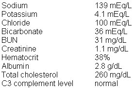
An 8-year-old boy is brought to the office by his mother because of recurrent episodes of "shortness of breath" and wheezing. These episodes typically occur when he is playing in the park with friends or when he is in the house at night. The symptoms are worst in the springtime and when he is watching television with his mother's boyfriend. The mother's boyfriend, who happens to smoke cigarettes, has been spending more and more time at the house, trying to bond with the patient. Pulmonary function tests show that the peak expiratory flow and forced respiratory volume per second are reduced during an attack and are normal during symptom-free intervals. Skin testing shows that he is allergic to grass and tree pollen, dust mites, animal dander, and a variety of other allergens. Laboratory studies show:

The most appropriate next step is to

  A. administer immunotherapy against identified allergens
  B. advise him to avoid all exercise
  C. advise him to try to avoid respiratory irritants, especially cigarette smoke
  D. advise the patient's mother to use a humidifier and air cleaners at home
  E. prescribe inhaled sodium cromoglycate, oral corticosteroids, and oral theophylline
Explanation:

The correct answer is C. This patient has asthma, and the most crucial step in the management of asthma is avoidance of the triggering factors, e.g., allergens. Unfortunately, it is difficult to avoid specific types of allergens, such as pollens. Specific measures to eliminate or reduce exposure to dust mites and animal dander at home lead to a reduced frequency of attacks and hospitalization rates. Regardless of the allergens involved, elimination of respiratory irritants, especially cigarette smoke, is of crucial importance. The bronchial tree of asthmatic patients is highly reactive to any form of chemical or physical irritation. Thus the avoidance of passive smoke is important. The mother should ask her boyfriend to go smoke outside alone if he needs to, but he should not be allowed to smoke in the house.

It is not practical to administer immunotherapy against identified allergens (choice A) in this case because he is allergic to multiple airborne allergens, and it seems like he is especially responsive to cigarette smoke. Immunotherapy is of some benefit when a single allergen is identified. The most important step is to try to reduce exposure to avoidable allergens (smoke).

Avoidance of all exercise (choice B) is not appropriate because even though exercise triggers asthmatic attacks in some patients, this does not seem to be his main trigger.

Humidifiers and air cleaners (choice D) at home is not the appropriate management. Humidifiers favor the growth of dust mites, and air cleaners have not been shown to be uniformly effective in getting rid of dust mites.

It is inappropriate to prescribe inhaled sodium cromoglycate, oral corticosteroids, and oral theophylline (choice E) for this patient because the fewest number of drugs at the lowest effective doses should be used. Typically, a one drug regimen (a bronchodilator or an inhaled corticosteroid) for mild to moderate asthma or two drugs for more severe cases is sufficient to control asthma exacerbations. Oral corticosteroids are indicated in cases of severe asthma and are therefore, not for this patient.


You have been following a 12-year-old girl who was diagnosed with autoimmune thrombocytopenic purpura of childhood (childhood ITP) 1 year ago following a viral illness. She has continued to have thrombocytopenia despite medical therapy. She recently received prednisone for 2 weeks followed by 2 days of intravenous immune globulin therapy. Her platelet count recently dropped below 20,000/mm3 requiring platelet transfusion and she repeatedly presents with diffuse petechiae and epistaxis. You and your colleagues decide that a splenectomy is the next step in treatment due to her persistent and dangerously low platelet count. Following the splenectomy and an uncomplicated postoperative course, she returns to your clinic for follow up. The thrombocytopenia has resolved and she has clinically improved. The most appropriate next step in this patient's management includes

  A. a 1-month course of penicillin prophylaxis and influenza vaccine
  B. a 1-month course of prednisone with concomitant IVIG therapy
  C. permanent penicillin prophylaxis, pneumococcus vaccine, and H. influenza vaccine
  D. permanent prednisone therapy and influenza vaccine
  E. a 2–week course of prednisone therapy
Explanation:

The correct answer is C. Autoimmune thrombocytopenic purpura of childhood (childhood ITP) is a disorder that usually occurs after a viral illness. The pathophysiology involves antibody (IgG or IgM) binding to platelets. These antibody- coated platelets are subsequently destroyed in the spleen. Thrombocytopenia ensues and most often resolves spontaneously within 6 months. Persistent thrombocytopenia is treated with 2 weeks of prednisone 2-4 mg/kg/day or IVIG 1g/kg/day. Cases refractory to medical therapy in which severe thrombocytopenia persists are treated with splenectomy to prevent further destruction of platelets. An extremely important aspect of management of the asplenic patient includes permanent penicillin prophylaxis in addition to pneumococcal and Haemophilus influenza vaccines. These measures decrease the risk of morbidity and mortality associated with overwhelming sepsis by encapsulated organisms in asplenic patients.

One month of penicillin prophylaxis and influenza vaccine (choice A) is an inappropriate choice. Patients should remain on penicillin prophylaxis due to the continued risk of infection. Asplenic patients are susceptible to infection with encapsulated organisms including Pneumococcus and H. influenza, thus vaccination against influenza is unnecessary.

One month of prednisone with concomitant IVIG therapy (choice B) is not indicated at this point. This patient has already failed these therapies and her condition has improved after the splenectomy.

Permanent prednisone therapy and influenza vaccine (choice D) is an inappropriate choice. Permanent prednisone would act to chronically suppress this patient's immune system making her more susceptible to an infection. Once again, influenza vaccination is not warranted in asplenic patients.

Two weeks of prednisone therapy (choice E) is inappropriate at this point. As stated before, prednisone therapy has already failed and the patient has improved post splenectomy.


A 45-year-old man comes to the office because of the progressive onset of difficulty in breathing during exercise. He reports that he has also felt more tired than usual lately. He denies any cough or chest pain. He smokes a pack of cigarettes a day and admits to heavy alcohol use over the past 3 years. His temperature is 37 C (98.6 F), blood pressure is 110/70 mm Hg, pulse is 105/min, and respirations are 18/min. Physical examination is remarkable for a third heart sound. A chest x-ray shows moderate enlargement of all 4 cardiac chambers and a small amount of pulmonary edema. An electrocardiogram shows sinus tachycardia and low voltage with nonspecific ST and T wave abnormalities. An echocardiogram shows mild dilatation of all chambers. At this time the most correct statement about his condition is:

  A. Cardiac catheterization will show increased cardiac output
  B. Ceasing alcohol consumption may reverse the course of his disease
  C. Immediate cardiac transplantation is indicated
  D. Spontaneous improvement occurs in 50% of patients with this disease
  E. Systemic embolization is not associated with his disease
Explanation:

The correct answer is B. This patient has dilated cardiomyopathy, which in this case is most likely due to his heavy alcohol consumption. Ceasing alcohol consumption may reverse his disease or at least halt its progression. Other treatments include salt restriction, diuretics, ACE inhibitors, digitalis, and the consideration of anticoagulation to decrease the risk of embolization.

Cardiac catheterization will show decreased cardiac output, not increased cardiac output (choice A) because of systolic dysfunction.

Immediate cardiac transplantation (choice C) is usually only indicated for advanced disease that is refractory to medical therapy.

Spontaneous improvement occurs in 50% of patients with this disease (choice D) is incorrect. While most people with dilated cardiomyopathy suffer progressive heart failure and death, about 25% of patients have a spontaneous improvement or stabilization.

Because of systolic dysfunction and the development of mural thrombi, systemic embolization is a serious concern for patients with dilated cardiomyopathy and therefore anticoagulation is often recommended. Systemic embolization is not associated with his disease (choice E) is incorrect.


A 30-year-old woman comes to the office for a periodic health maintenance examination. She has no complaints at this time. She works as a sales manager of a department store, goes to an aerobic exercise class after work, 4 times a week, drinks a glass of wine every 3-5 days, and does not smoke cigarettes. She tells you she recently stopped taking her oral contraceptive pills because she and her husband want to start trying to have a baby soon. Her menstrual periods come at regular 28-day intervals and typically last for 6 days. Her last menstrual period was 10 days ago. Her blood pressure is 110/70 mm Hg and pulse is 60/min. Physical examination is unremarkable. A pelvic examination is unremarkable. You decide to perform a Pap smear because you see that her last one was 2 years ago and was normal, as always. The most appropriate next step is to

  A. do a urine pregnancy test
  B. obtain chlamydial and gonorrheal cultures
  C. perform a clinical breast examination
  D. recommend a daily multivitamin with folic acid
  E. test her for hepatitis B surface antigen
Explanation:

The correct answer is D. The United Stated Preventive Services Task Force (USPSTF) recommends that all women planning to become pregnant take a daily multivitamin supplement containing folic acid. It is recommended that they start taking it at least 1 month prior to conception and continue through the first trimester to reduce the risk of neural tube defects. According to the USPSTF, taking a daily multivitamin containing folic acid is also recommended for all women capable of becoming pregnant. This is to reduce the risk of neural tube defects in unplanned pregnancies.

A urine pregnancy test (choice A) is unnecessary at time because this patient's last menstrual period was only 10 days ago and she has not missed a period. It is very unlikely that she is pregnant at this time.

It is unnecessary to obtain chlamydial and gonorrheal cultures (choice B) because this patient is asymptomatic and is not considered at high risk for these infections. The USPSTF recommends that the routine screening of asymptomatic persons for these infections should typically be limited to sexually active adolescents and people that fall into high-risk groups, including those with a prior sexually transmitted disease, multiple partners, inconsistent barrier contraception usage, being unmarried, and being under 25 years of age. High-risk pregnant women should be screened. They say that the routine screening of asymptomatic adults for chlamydia and gonorrhea is not recommended.

The USPSTF concludes that there is insufficient evidence to recommend for or against routine clinical breast examinations (choice C) alone to screen for breast cancer. They recommend screening mammography, with or without clinical breast examination, every 1–2 years for women aged 40 and older. According to these guidelines, this asymptomatic 30-year-old woman does not need a CBE at this time.

The USPSTF recommends that pregnant women be tested for hepatitis B surface antigen (choice E) at their first prenatal visit. Since she is most likely not pregnant yet (her last menstrual period was 10 days ago), this test is unnecessary at this time.


A 54-year-old woman comes to the office for a follow-up examination 6 weeks after starting lovastatin for elevated cholesterol. She has been a patient of yours for years, and you have treated her for hypertension, an episode of gout, and anemia caused by uterine leiomyomas that were treated with a hysterectomy 5 years ago. She has no complaints at this time and is in a rush to pick up her children from a soccer game. Her blood pressure is 130/80 mm Hg and her pulse is 65/min. Physical examination is unremarkable. The most appropriate course of action is to order cholesterol levels and to

  A. let her go pick up her children
  B. obtain uric acid levels
  C. order creatine kinase levels
  D. order liver function tests
  E. remind her to call you if she develops any side effects from the medication
Explanation:

The correct answer is D. Abnormal liver function is one of the main side effects associated with HMG-CoA reductase inhibitors, which are one of the most common classes of drugs prescribed for elevated cholesterol. While this side effect is relatively uncommon (1-2%), it can be very serious. It is important to monitor cholesterol and liver function tests at 6 weeks and 12 weeks after the initiation of therapy or when the dosage is increased, and then every 4-6 months.

It is important to order liver function tests and to remind her to call you if she develops any side effects from the medication, before you let her go pick up her children (choice A).

There is no apparent reason to measure uric acid levels (choice B) in this patient at this time. Niacin, not HMG CoA reductase inhibitors, is associated with elevated uric acid levels. Even though this patient has a history of gout, she does not have symptoms now and she is specifically at the office for a follow-up examination after starting lovastatin.

Myositis is one of the other side effects of HMG CoA reductase inhibitors, but this too is relatively uncommon. Myositis occurs more frequently when combined with other cholesterol lowering agents. Discontinuation of the medication is important if these symptoms occur because rhabdomyolysis with renal failure and death can occur. The routine measurement of creatine kinase levels (choice C) is not recommended. It is usually done when patients complain of muscle aches and cramps.

While it is important to remind her to call you if she develops any side effects from the medication (choice E), such as muscle aches, you first need to order liver function tests.


A 16-year-old girl is brought to the office for a gynecologic examination a week after her first sexual experience. You have been her physician since she was a baby, performing camp and school physical examinations, and providing immunizations. You know that she has never had a pelvic examination before, and can tell that she is very nervous. She is sitting on the examination table, biting her cuticles and looking down at the floor. After you ask her mother to leave the room, the patient tells you that she and her boyfriend had sexual intercourse for the first time, and that they used condoms and spermicidal lubricant as contraception. You inquire about any other pertinent issues, and she tells you that she has no specific concerns, and that everything else is going pretty well. She lies down on the examination table and places her heels on the foot rests. You drape a sheet over her knees and sit down at the end of the table. You can see that she is clenching her teeth. The most appropriate statement at this time is:

  A. "How many times have you and your boyfriend had sexual intercourse?"
  B. "Please try to relax as much as you can. I know that is easier said than done."
  C. "Relax! This is not going to hurt."
  D. "Why are you clenching your teeth? I'm not going to hurt you."
  E. "Why are you so nervous? You've already had sex so this shouldn't hurt."
Explanation:

The correct answer is B. It is important to realize that a pelvic examination is an uncomfortable procedure for women to go through, and the position may make them feel nervous, embarrassed, and helpless. Helping women to relax and feel comfortable makes the whole experience easier for both the physician and the patient. The best statement at this time is, "Please try to relax as much as you can. I know that is easier said than done." It may help her relax, and also make her realize that you understand that this is a difficult situation.

"How many times have you and your boyfriend had sexual intercourse?" (choice A) is not a relevant question, because no matter what the answer is, it will not change your management. Also, you need to recognize that her "clenched teeth" probably means that she is nervous, and needs reassurance at this time.

"Relax! This is not going to hurt." (choice C) is almost like a command, and it may make her even more nervous. "Please try to relax as much as you can. I know that is easier said than done," is a much better way to get a similar message across.

"Why are you clenching your teeth? I'm not going to hurt you," (choice D) is inappropriate because it may make her even more nervous. Also, "why" questions like this one have the possibility of making the patient defensive and uncomfortable.

"Why are you so nervous? You've already had sex so this shouldn't hurt." (choice E) is completely inappropriate, because you should realize that a pelvic examination is an uncomfortable situation, especially the first one, and your main goal at this time should be to help her relax. Also, the second part, "You've already had sex so this shouldn't hurt," is a terrible thing to say, because you may think that you are being friendly or funny, but your job is to be a professional, and not make inappropriate statements. It seems a bit judgmental and may be seen as sexual harassment. Another point is that you do not know if this is going to hurt her, whether or not she has had sex already. It is best to avoid these types of statements all together, and say something that will help put her at ease.


A 61-year-old woman comes to the office because of lower and upper extremity swelling. She has a long history of hypertension, hyperlipidemia, and gout that have been very well controlled. She is an active woman who works as a fashion store manager. She takes thiazide, mevastatin, and allopurinol daily. Over the past few weeks, she has noticed increasing swelling of her feet and her hands. Her feet have gotten so swollen that this morning she was unable to put her shoes on. Her temperature is 37 C (98.6 F), blood pressure is 180/70 mm Hg, pulse is 72/min, and respirations are 12/min. Blood chemistries are remarkable for a BUN of 40 mg/dL and a creatinine of 1.8 mg/dL. A urine dipstick is positive for protein. A 24-hour urine test confirms 4gm of protein. The most important intervention at this time is to

  A. add a loop diuretic
  B. increase thiazide dosage
  C. initiate ACE inhibitor therapy
  D. recommend a high protein diet
  E. start hemodialysis
Explanation:

The correct answer is C. The nephrotic syndrome is defined by a urinary protein level exceeding 3.5 g per 1.73 m2 of body-surface area per day. Diabetic nephropathy is the most common cause of nephrotic proteinuria. Five primary glomerular diseases account for the great majority of cases of the nephrotic syndrome in persons who do not have diabetes. In adults, the most common cause is membranous glomerulonephropathy. A common clinical triad of the nephrotic syndrome is hypertension, hyperlipidemia, and proteinuria. Although the exact mechanism whereby edema formation occurs in these patients is uncertain, the loss of urinary protein leads to total body edema formation. Regardless of the magnitude of the urinary protein loss, initiating ACE inhibitor therapy has been shown to be beneficial in terms of both decreasing the urinary protein content and prolonging survival.

Adding a loop diuretic (choice A) or increasing the thiazide dosage (choice B) fails to address the etiology of the edema. This patient has new onset edema and simply trying to manage the symptom by increasing an existing diuretic dosage or adding a second diuretic class agent fails to address the underlying etiology of this patient's edema.

There is no role for a high-protein diet (choice D) in managing these patients. Contrary to previously held opinions that the protein needed to be replaced by high-protein diets, it is now clear that such diets exacerbate kidney damage and accelerate protein loss. Low-protein diets are recommended for these patients.

There is no indication for hemodialysis (choice E) at this time. The five indications for HD are refractory hyperkalemia, volume overload, acidosis, uremia, or uremic pericarditis.


A 15-year-old boy comes to the office for a physical examination before going to summer camp. He says that he has a tender "lump" in his right breast that he noticed a couple of months ago. He is very concerned because he is going to a new camp and he is worried that he is going to have to change clothes in front of other boys in his bunk. He thinks that the other boys are going to see it and make fun of his "breast". There are tears streaming down his face as he tells you this. Physical examination shows a 1.5-cm, tender, palpable mass symmetrically distributed beneath the right areola. There is no discharge from the right nipple. The left breast is unremarkable. The remainder of the physical examination shows a small amount of dark, curling pubic hair and open and closed comedones on his cheeks, forehead, and back. The most appropriate next step is to

  A. order liver function tests
  B. perform ultrasonography of the breast
  C. prescribe testosterone to give him a more "masculine" body-type
  D. reassure him that this is common and no further testing is indicated
  E. refer him for a fine needle aspiration
  F. schedule him for a mammography
Explanation:

The correct answer is D. Gynecomastia (puberal hypertrophy) is very common in adolescents during puberty and is frequently asymmetric and tender. It is important to reassure this patient that it affects approximately 50–60% of adolescent boys at around Tanner stage III. It usually regresses before age 20. Surgery is rarely indicated. The open and closed comedones (also known as "blackheads" and "whitehead") are part of acne vulgaris, which is another common problem that affects adolescents.

Evaluation for liver disease (choice A) is unnecessary at this point because gynecomastia is very common during adolescence. He has no other signs of liver disease.

An ultrasound (choice B) is often used to distinguish cystic from solid masses. It is not indicated at this time in this case because this patient most likely has puberal hypertrophy of the breast (gynecomastia).

It is completely inappropriate to prescribe testosterone to give him a more "masculine" body-type (choice C) because testosterone often causes gynecomastia. Testosterone is converted to estradiol in extraglandular tissues and leads to feminization.

Fine needle aspiration (choice E) and mammography (choice F) are used to evaluate a dominant breast mass or possibly gynecomastia in a patient who is not going through puberty, has a negative drug history, or a rapidly growing, large (>4cm) mass. This patient's gynecomastia is most likely due to puberty, making further work-up too aggressive at this time.


A 42-year-old college professor comes into the office complaining of a "flare of acne" in recent years associated with increased flushing. On further questioning, she tells you her face becomes unbearably red when she drinks coffee or wine at dinner parties, goes out for short hikes on weekends, or has spicy Thai food. She has also noticed an increasing number of enlarged veins on her cheeks and nose. She is currently on ibuprofen for low back pain but denies any other medications. She has no known allergies. Face and neck examination shows telangiectasias on the tip of her nose, bilateral cheeks, and forehead as well as pustules and inflammatory papules over cheeks and glabella. There is no involvement of the neck. She should be told that she most likely has

  A. acne rosacea and should avoid exacerbating factors and use oral tetracycline and topical metronidazole
  B. contact allergic dermatitis to her makeup and she should switch to a different line of makeup
  C. cystic acne and should take isotretinoin after establishing that she is definitely not pregnant
  D. drug allergy to ibuprofen and she should discontinue its use
  E. perioral dermatitis and she should use oral minocycline and topical tretinoin cream
Explanation:

The correct answer is A. Acne rosacea is a chronic inflammatory skin condition that has a predisposition to affect the central "flush" areas of the face, the central cheeks, nose, brow, and chin. This condition most frequently takes the form of flushing erythema which appears to be triggered by the consumption of alcohol, spicy food, or hot foods and beverages. Environmental factors that exacerbate flushing are sun exposure, heat, cold, and wind. The second main component to this disorder is acneiform papules and pustules, furuncles, and cysts. Treatment of rosacea includes avoiding exogenous factors, as mentioned above, and the use of a variety of topical and oral antibiotics. The most common topical antibiotic used by dermatologists is metronidazole gel, which is applied to affected areas twice a day. If the patient experiences moderate to severe inflammatory papules and pustules, the addition of an oral antibiotic is indicated, most commonly one in the tetracycline family.

Contact allergic dermatitis (choice B) is incorrect because usually contact dermatitis to makeup will result in generalized erythema over the surface that comes in contact with the culprit agent as well as numerous small, pinpoint vesicles. It is not usually exacerbated by foods, drinks, or the sun.

Cystic acne (choice C) is incorrect because the woman is described as having inflammatory papules and pustules, not deep-seated cysts. Also, cystic acne is usually not triggered by foods or drinks. Also, this is the classic description of acne rosacea.

Drug allergy to ibuprofen (choice D) is incorrect because drug allergies tend to involve the entire body, beginning at trunk then spreading to the extremities with the face being affected last.

Perioral dermatitis (choice E) is incorrect because perioral dermatitis is a discrete eruption consisting of grouped erythematous papules, vesicles, or pustules located around the mouth with marked sparing of the skin immediately surrounding the vermilion border. It often occurs on the nasolabial folds, on the chin, and around the eyelids.


A 71-year-old man with mild hypertension and high cholesterol comes to the office complaining of 2 weeks of intermittent vertigo with each episode lasting about 2-4 hours. He also reports hearing a low frequency buzzing, which is constant but waxes and wanes in intensity. He tells you that over this time he has been having trouble hearing while in noisy areas such as in restaurants or temple gatherings. Physical examination is normal. Vertigo is not exacerbated by changes in head position. The most appropriate management of this patient is to

  A. begin diazepam therapy
  B. begin hydrochlorothiazide therapy
  C. begin meclizine therapy
  D. begin scopolamine therapy
  E. recommend physical therapy
Explanation:

The correct answer is B. This patient has Meniere's syndrome (endolymphatic hydrops). Meniere's syndrome is characterized but intermittent vertigo lasting about 1-8 hours with associated hearing loss, aural pressure, and tinnitus. Symptoms tend to wax and wane. It differs from benign positional vertigo in that the symptoms do not necessarily worsen with positional changes. In addition, positional vertigo doesn't have the same associated factors as Meniere's syndrome does. The treatment is a low salt diet and HCTZ. If the patient is resistant to medical therapy, surgery to decompress the endolymphatic sac can be a last resort.

Diazepam (choice A) is a useful treatment to ablate an acute episode of acute vertigo but is not a first line therapy for Meniere's syndrome.

Meclizine (choice C) is an antihistamine, which is useful in the management of less severe attacks of vertigo.

Scopolamine (choice D) is sometimes used in a transdermal preparation to be beneficial in the management of chronic vertigo. The anticholinergic side effects can limit its usefulness.

Physical therapy (choice E) is becoming more important in the management of vertigo. It is thought to help the enhance CNS ability to compensate for labyrinthine dysfunction. Recently, use of specific head maneuvers has been incorporated into the management of vertigo.


A 27-year-old woman, who had a tuberculosis skin test placed 3 days earlier because of a cough, night sweats, and weight loss, comes back to the office to show you the results. There is a 16mm area of induration at this injection site. She has never had any induration on past tuberculosis skin tests. A chest x-ray shows hilar adenopathy, upper lobe infiltrates, and a cavity. You obtain a sputum sample culture for acid-fast bacilli. You discuss this condition with her and she is very concerned about the health of the asymptomatic newborn baby boy that she adopted 3 months ago. The cultures return a week later and are positive for acid-fast bacilli. She is given appropriate treatment and the board of health is notified. The patient's husband brings the newborn into the office for evaluation. The tuberculosis skin test and chest x-ray that are performed on the baby, are both negative. At this time you should

  A. do nothing
  B. obtain a sputum sample by bronchoscopy from the newborn
  C. prescribe a 6-month course of isoniazid, rifampin, pyrazinamide, and ethambutol for the newborn
  D. prescribe a 3-month course of isoniazid for the newborn
  E. reevaluate the newborn in 3 months
Explanation:

The correct answer is D. The mother has tuberculosis disease and the newborn has no evidence of infection or disease. According to the American Academy of Pediatrics and the Report of the Committee on Infectious Diseases, a newborn infant should be given prophylaxis with isoniazid if a recent contact has active disease. This is true even if they have a negative tuberculosis skin test (TST) and chest x-ray. The newborn should be reevaluated with a skin test in 3 months and if negative the isoniazid can be discontinued, but if positive the isoniazid should be continued for 6 more months (9 months total).

The American Academy of Pediatrics recommends that household contacts (younger than 4 years old) of patients with active disease should be treated if they have a negative skin test and chest x-ray. Doing nothing (choice A) and reevaluating the newborn in 3 months (choice E) are incorrect.

If the TST and the chest x-ray are negative and the patient is asymptomatic, it is unnecessary to obtain a sputum sample by bronchoscopy from the baby (choice B).

Isoniazid, rifampin, pyrazinamide, and ethambutol (choice C) are treatment for tuberculosis disease (positive TST, signs and symptoms of disease, and an abnormal chest x-ray and sputum). The newborn does not have the disease and should not be given all of these drugs.


A 39-year-old man with no significant past medical history comes to the office because of "red urine." He tells you that he has been well over the past few years, but has occasionally noticed hematuria in the morning. His family history is unremarkable. He denies any tobacco or intravenous drug use. His blood pressure is 180/100 mm Hg and pulse is 70/min. Physical examination shows clear lungs, normal heart sounds without murmurs, and 3+ pitting edema of his lower extremities bilaterally. Laboratory studies show:

The most likely diagnosis is

  A. focal segmental glomerulosclerosis
  B. IgA nephropathy
  C. membranoproliferative glomerulonephritis
  D. rapidly progressive glomerulonephritis
  E. systemic lupus erythematosus
Explanation:

The correct answer is B. IgA nephropathy, also called Berger disease, is a major cause of recurrent glomerular hematuria. The hematuria typically lasts for a few days, and then subsides, only to recur every few months. The total picture is that of a nephrotic syndrome, with hyperlipidemia, proteinuria, and hypoalbuminemia. Most patients have an initial benign course, but up to 50% develop chronic renal failure by 20 years.

Focal segmental glomerulosclerosis (choice A) is a rare cause of idiopathic glomerular disease, usually characterized by hypertension, hematuria, renal insufficiency, and nephrosis. Compared to Berger disease, this entity in its primary form is quite rare.

Membranoproliferative glomerulonephritis (choice C) exhibits a variety of presentations ranging from asymptomatic proteinuria to full nephrotic syndrome. The diagnosis should be suspected when renal disease is accompanied by low serum complement levels. Hepatitis C virus accounts for most of the cases of MPGN.

Rapidly progressive glomerulonephritis (choice D) presents with rapidly declining renal function, an active urinary sediment including RBC casts, as well as oliguria or anuria.

Systemic lupus erythematosus (choice E) may involve the kidney and can have a variety of renal presentations; from benign progressive azotemia to rapidly progressive nephritis. The kidneys are involved in nearly all cases of SLE, but rarely without involvement of other organs.


The mother of a 3-year-old patient of yours calls the office in the morning to report that her daughter woke up with a "red eye with a thick yellow discharge." You do not have any open appointments in the morning, and since the girl does not have severe pain or discomfort and has no change in vision, you schedule an appointment in the afternoon. You look over the patient's chart and note that she is generally a very healthy child who is developing normally. At this time you should tell the mother that:

  A. Careful hand washing does not affect the spread of this condition
  B. She should be excluded from child care until after your examination and approval for readmission
  C. She should take her daughter for a CT scan before the appointment to rule out orbital cellulitis
  D. Since the condition is most likely due to a self-limited virus, the appointment is for reassurance and not to administer therapy
  E. Topical corticosteroids are indicated if the patient has a herpetic dendritic ulcer
Explanation:

The correct answer is B. This patient most likely has conjunctivitis due to a bacterial infection. Conjunctivitis is a common condition in children that is usually bacterial or viral. Bacterial infections are usually purulent, while viral infections are not. Topical antibiotic therapy is typically given for bacterial infections. Conjunctivitis is contagious and it usually spreads by direct contact. Respiratory spread may occur. Children should be excluded from childcare settings until they are examined, given treatment if indicated, and approved for readmission.

It is incorrect to tell the mother that careful hand washing does not affect the spread of this condition (choice A) because the spread of infection is minimized by hand washing.

It is inappropriate to tell the mother that she should take her daughter for a CT scan before the appointment to rule out orbital cellulitis (choice C). A CT scan is used to evaluate a patient with suspected orbital cellulitis, which presents with proptosis, decreased visual acuity, and pain with movement. It is unlikely that the patient in this case has orbital cellulitis, and if the symptoms that the mother reported were consistent with orbital cellulitis, you would want to evaluate her before sending her for diagnostic studies.

Since the condition is most likely due to a self-limited virus, the appointment is for reassurance and not to administer therapy (choice D) is incorrect. The "thick yellow discharge" that the mother described is consistent with a bacterial infection, not a viral infection. The appointment is for evaluation and the administration of antibiotic therapy if indicated.

Topical corticosteroids are indicated if the patient has a herpetic dendritic ulcer (choice E) is incorrect. Corticosteroids are contraindicated in patients with a herpetic dendritic ulcer because they can worsen the condition. Individuals with a dendritic ulcer often have a painful, red eye, with visual blurring. Discharge may be present.


A 77-year-old man comes to the office with his wife because of "walking difficulties." He says that over the past 5 months he has noticed that when he walks or stands for longer than 15 minutes he gets pain and weakness in his thighs. The pain is usually relieved by sitting. Within the past 1-2 years he began to get a "discomfort" in the anterolateral thighs, more in the right lower extremity than the left. He also gets a pain in his right hip, which radiates down to just below his knee. He denies ever having any calf pain. He urinates 2-3 times per night and will lose 1 or 2 drops of urine if he cannot make it to the bathroom in time. His wife has notice that he has a tendency to stand with his knees slightly bent rather than straight legged. He tells you that 16 years ago he began to feel "unsteady on his feet." He did not fall or experience pain at that time, but he had "pins and needles feelings" in his fingers and feet and "lost the feeling of his feet being attached to the ground." He saw 2 different doctors at that time, had a myelogram, and was diagnosed with C4-5 damage. He underwent C4-5 intercervical discectomy and osteophyte removal. After the surgery he wore a neck brace for several months and the symptoms remained stable. He noticed that his knee reflexes were stronger after the surgery. 4-5 years ago he began to notice that his right knee would buckle. This resulted in 2-3 falls over a 1-year period. He saw a neurologist who prescribed physical therapy and a cervical collar to be worn at night. He did well and stopped wearing the collar about 1 year ago. Physical examination shows weak, but palpable distal pulses, moderately limited neck range of motion, mild weakness of the deltoids and biceps bilaterally, mild weakness of hamstrings and extensor hallucis longus bilaterally, and a normal sensory exam. He has brisk symmetric deep tendon reflexes and down-going toes bilaterally. Tests of coordination are normal and his gait is normo-based and steady but mildly spastic. Cranial nerve and mental status examinations are unremarkable. The most appropriate next step is to

  A. order an MRI of the brain
  B. order an MRI of the cervical spine
  C. order an MRI of the lumbar spine
  D. perform electromyography and nerve conduction study
  E. send him for an angiogram of the lower extremities
Explanation:

The correct answer is C. The history is typical of that for neurogenic claudication, which is caused by lumbar stenosis. The lower extremity weakness is also typical of bilateral L4/L5 radiculopathies. You would want to do an MRI to confirm the diagnosis and make sure that other pathology such as a spinal tumor was not responsible.

There is no reason to suspect brain pathology in this case (choice A). If there were cranial nerve or mental status abnormalities it should be considered.

His gait disorder is being caused primarily by pain, which is secondary to neurogenic claudication of the lumbar spine. He has known cervical spine problems, which may be contributing by impairing balance. It would also cause weakness of deltoids and biceps by C5 root compression. He may need a cervical spine MRI (choice B) at some point, but it is not primarily causing his walking troubles.

There is no indication for electromyography or nerve conduction (choice D) as the history and exam are sufficient to make this diagnosis. An MRI should be ordered to confirm the diagnosis and rule out other pathology.

An angiogram of the lower extremities might be indicated if you suspected vascular claudication (choice E). More commonly this would involve the calves and distal pulses would not be palpable.


A 27-year-old woman comes to the office for a periodic health maintenance examination. She is a healthy patient with great habits. She exercises 3 times a week, takes 1500 mg of calcium daily, does not drink or smoke, and eats a low-fat, high fiber diet. Her last Pap smear, which was 2 years ago, was normal, and her blood pressure, body mass index, and non-fasting cholesterol have all been stable. A focused physical examination is unremarkable. You decide that a complete pelvic examination is indicated at this appointment. The external genitalia, vagina, and cervix appear normal. The most appropriate next step is to

  A. do a bimanual examination
  B. obtain chlamydia and gonococcal cultures
  C. obtain the endocervical specimen
  D. obtain the ectocervical specimen
  E. put lubricant on the Pap smear spatula
Explanation:

The correct answer is D. When performing a complete pelvic examination, the Pap smear is the first specimen to collect to avoid contamination. Both endocervical and ectocervical specimens should be obtained during a Pap smear. However, the ectocervix should be sampled first, to avoid contamination with the endocervical cells, and therefore, get an accurate sample. Lubricant should not be used until after collecting cervical cells with a Pap smear.

A bimanual examination (choice A), should be performed after the Pap smear. This is to make sure that the Pap smear specimen is not contaminated by lubricants and cells, (or bacteria), from other areas.

If you are going to obtain chlamydia and gonococcal cultures, (choice B), it should be done after the Pap smear. This is to make sure that the Pap smear specimen is not contaminated by lubricants and cells, (or bacteria) from other areas. Routine screening for chlamydia in asymptomatic individuals is recommended for high risk women, and sexually active women, and adolescents age 25 and younger. As of 2001, the USPSTF makes no recommendation for or against the routine screening of asymptomatic low-risk women in the general population for chlamydial infection. Routine screening for gonorrhea in asymptomatic individuals is recommended for high risk women.

The endocervical specimen (choice C), should be obtained after the ectocervical specimen. When performing a Pap smear, it is important to obtain an accurate sample and to be certain as to exactly where the cells are taken from. If you obtain the endocervical cells first, there is a chance, that some of these cells will be deposited on the exocervix, and then when the ectocervical sample is taken, the endocervical cells, with be mixed with the ectocervical cells and the pathology reading will not be accurate, (as to the site).

A lubricant (choice E), should not be used on the Pap smear spatula. It may contaminate the specimen.


An 8-month-old infant, who you have been taking care of since he was born at the local community hospital, is brought to the office because of constipation. The father, a "stay-at-home dad," tells you that she has been having 1 bowel movement every 3-4 days, and that the stool is always very hard. He says that she is doing very well otherwise; she is a very happy and easy little girl. She is fed primarily infant formula and he is starting to introduce solid foods. He says that he is concerned because he remembers always having to change "very dirty" diapers for both of his other children, at least twice a day. Physical examination is unremarkable. A rectal examination shows guaiac negative brown stool. The most appropriate next step is to

  A. advise him to give her mineral oil 3 times a day until she is "regular"
  B. advise him to give her prune juice or pear juice
  C. determine thyroid-stimulating hormone levels
  D. order a barium enema
  E. order rectal manometry and a rectal biopsy
  F. reassure him that all infants have different bowel habits
Explanation:

The correct answer is B. Constipation is a common problem in formula-fed infants, and it is best treated by increasing the amount of fluids in the diet, especially with fruit juices that contain sorbitol, such as prune and pear, which help to relieve constipation. It is often caused by a diet that is too low in fluids or deficient of bulk.

It is not recommended to give mineral oil (choice A) to infants because of the risk of aspiration and severe lipoid pneumonia.

Determining TSH levels (choice C) is not appropriate at this time because this is a common problem in formula-fed infants that is related to diet. Hypothyroidism is associated with constipation, however, newborns born in United States hospitals are routinely screened for this disorder, which can also lead to mental retardation. It is unlikely that she has hypothyroidism.

A barium enema (choice D), rectal manometry and a rectal biopsy (choice E) are all part of the evaluation of patients with suspected Hirschprung's disease, which is a lack of ganglion cells in the distal colon. It is very unlikely that this infant has this relatively rare (1 in 5000) disease that is often suspected if a newborn does not have stool in the first 24 hours of life. Infants with this disease often have a distended abdomen and a rectum that is devoid of stool. It is more likely that this formula-fed infant has diet-related constipation.

Reassurance (choice F) is incorrect in this case because formula-fed infants this age should be having 1-2 bowel movements a day. You can reassure him that this is very common and she will most likely be fine once they adjust her diet.


A 40-year-old man comes to the office complaining of a 3-day history of midsternal chest pain, non-radiating that is worse with inspiration and relieved by sitting forward. He has no past medical history, is on no medications, does not smoke, and has no known drug allergies. He leads an active lifestyle, and had been running about 10 miles a week without problem until a week ago when he developed a “viral syndrome.” His temperature is 38.4 C (100 F), blood pressure is 130/70 mm Hg, pulse is 100/min and regular, and respiratory rate is 20/min. He has a high pitched, grating sound that can be auscultated throughout the cardiac cycle over his precordium. An electrocardiogram shows diffuse ST elevation, diffuse PR depression with PR elevation in lead aVR. The most likely diagnosis is

  A. angina
  B. myocardial infarction
  C. pericarditis
  D. pneumonia
  E. pulmonary embolism
Explanation:

The correct is answer is C. A chest pain that is pleuritic and improves with sitting up and leaning forward is a classic description of the chest pain associated with pericarditis, as is the precordial rub auscultated on physical exam. Diffuse ST elevations with diffuse PR depressions and PR elevation in lead aVR is also the classic description of the EKG findings associated with pericarditis.

Angina (choice A) or myocardial infarction (choice B) are less likely given that the patient has no risk for coronary artery disease (hypertension, hyperlipidemia, diabetes, tobacco). He also describes a very active lifestyle without symptoms, suggestive of no cardiac pathology prior to the onset of these symptoms. The ST changes on EKG, that alone would suggest ischemia, become more consistent with pericarditis since they are associated with PR segment changes that are classically associated with this particular disease process.

Pneumonia (choice D) is unlikely since there is no history of cough or dyspnea, and there is no mention of findings on the chest exam consistent with such a process (e.g., findings suggesting a consolidative process such as decreased breath sounds, increased fremitus, and egophony over the affected area).

Pulmonary embolism (choice E) is less likely, given that there are no apparent risk factors such as a history of hypercoagulability or poor activity. The EKG findings are also not consistent with a pulmonary embolism, where one would classically see an S wave in lead I, Q wave and T wave inversion in lead III, or ST-T wave changes in leads V1 through V4.


A 57-year-old man comes to the office because he has been feeling tired and sad for the past 3 months. He has had difficulty concentrating at work and besides a general "lack of energy," he has noticed that he is having a little trouble with his memory. He denies any recent change in weight, or thoughts of death. He works as a hospital administrator, has been married for 29 years, and has 2 kids. He has no significant past medical history or family history, and does not take any medications. His temperature is 37.0 C (98.6 F), blood pressure is 110/80 mm Hg, pulse is 70/min, and respirations are 16/min. Physical examination is unremarkable. The most appropriate next step is to

  A. advise him to take St. John's Wort and get plenty of exercise and exposure to daylight
  B. obtain thyroid function tests, a biochemical profile, and a complete blood count
  C. order a CT scan of the abdomen
  D. prescribe fluoxetine and ask him to return for reexamination in 2 weeks
  E. refer him to a psychiatrist
Explanation:

The correct answer is B. This patient is presenting with symptoms of depression. However, major organ disease must be ruled out before the diagnosis is made. Laboratory studies should be ordered, including thyroid function tests, a biochemical profile, and a complete blood count. Diseases such as hypothyroidism, anemia, diabetes mellitus, and disorders of the kidney, liver, and cardiovascular systems are all associated with depressive symptoms.

Some patients say that taking St. John's Wort (SJW) and getting plenty of exercise and exposure to daylight (choice A) are helpful in treating depressive symptoms, but before you make any recommendations for his symptoms, he should be evaluated to rule out an organic disease. SJW is an herbal medication that inhibits the uptake of serotonin, dopamine, and norepinephrine. There is some clinical data to support its use for depression, but this is not widely accepted.

It is inappropriate to order a CT scan of the abdomen (choice C) to evaluate this patient's depressive symptoms because it is too expensive and most likely unnecessary. Pancreatic cancer has been associated with depression, but this patient does not have any other symptoms that are suggestive of pancreatic cancer (jaundice, pain, weight loss). Laboratory studies are appropriate at this time.

After laboratory studies rule out any abnormalities, it is appropriate to prescribe fluoxetine and ask him to return for a reexamination in 2 weeks (choice D). It is not the next step at this time.

Referral to a psychiatrist (choice E) may be necessary in the future, but at this time laboratory studies should be ordered and if they are all normal, a trial of antidepressant therapy should be started. If he does not respond or if you (and the patient) think that he may benefit from mental health care, he should be referred.


An 18-month-old boy is brought to the office because his mother claims that he appears very pale but is otherwise acting normally. She tells you that he drinks approximately 48 ounces of whole milk per day. There is no history of anemia in the family. His temperature is 37.0 C (98.6 F), blood pressure is 80/50 mm Hg, pulse is 120/min, and respirations are 20/min. Physical examination shows pallor of the skin and mucous membranes. A 2/6 systolic ejection murmur is noted. Laboratory studies show:

The most appropriate initial management is to

  A. evaluate the family for anemias
  B. give folate, orally
  C. give vitamin B12, intramuscularly
  D. recommend that the patient cut down on the milk intake and start oral iron supplementation
  E. transfuse with whole blood
Explanation:

The correct answer is D. Iron deficiency is the most common nutritional deficiency in children ages 9-15 months. It can be due to the decreased availability of dietary iron, impaired absorption of iron secondary to frequent infections, or an increased requirement of iron for growth. Also, iron deficiency caused by high cow's milk intake is a relatively common problem in young children. Frequently, parents will continue giving large amounts of milk to their children because they are used to giving as much formula as the child wants during the first year of life. This causes a few problems. First, it will cause the child to eat less regular food and therefore not receive the vitamins and minerals necessary for growth. In addition, as seen in this case, iron deficiency can occur. Milk doesn't contain much iron and through mechanisms not well understood, it also inhibits iron absorption. The anemia can be profound in many cases and may require transfusion. In this case the signs of iron deficiency are seen in the very low MCV, high RDW, and anemia. Since this is a chronic process, the anemia is well tolerated by the child and therefore he is not symptomatic. Therefore he just requires iron supplementation and a decrease in the daily milk intake to around 24 ounces per day. With oral iron a prompt reticulocytosis and rise in hemoglobin and hematocrit will be seen.

At this point an expensive evaluation of the family for anemias (choice A) is not warranted because the source for the anemia seems apparent. If after a few months of iron and a decrease in milk intake the RBC indices and Hct do not improve, then further investigation in necessary.

Folate deficiency would cause a macrocytic anemia and is not a consequence of excessive cow's milk intake. Folate deficiency can be seen in children who are raised on goat milk. Giving folate (choice B) to this patient is inappropriate.

As with folate deficiency, B12 deficiency causes a macrocytic anemia that is not seen in this case and is not very common in young children with no other illnesses or deficiencies. Therefore giving B12 (choice C) to this patient is inappropriate.

Transfusion (choice E) is not warranted here because the patient, although pale, is asymptomatic. In addition, children are usually transfused with packed RBCs to restore hemoglobin not whole blood.

A 61-year-old man comes to the office complaining of joint stiffness and pain. He is otherwise healthy and enjoys an active lifestyle but reports that over the past few months his hands and knees have begun to ache as his day progresses and that they often become "tight" or "stiff". He has difficulty extending his legs completely and his hands, he finds, are less dextrous than a few years ago. His vital signs are normal. His knees show mild crepitus bilaterally with some tenderness to palpation of the left knee and his PIP joints of his fingers are mildly enlarged. A radiograph of his knees shows narrowing of the joint spaces bilaterally with subchondral sclerosis. At this time the most correct statement about this patient's condition is:

  A. A fixed daily dose of nonsteroidal anti-inflammatory drugs is indicated
  B. He requires knee replacement surgery for pain relief and increased mobility
  C. MRI of the knees is indicated for a more detailed joint evaluation
  D. Nonpharmacologic management and acetaminophen therapy should be initiated
  E. These are normal age-related changes to his joints
Explanation:

The correct answer is D. This patient has osteoarthritis that is a degeneration of the cartilage and hypertrophy of bone at the articular margins. Radiographs often show the characteristic joint-space narrowing, subchondral sclerosis and cyst formation, and marginal osteophytes. There is however minimal correlation between the severity of the radiographic findings and the clinical symptoms or functional limitations. This patient has mild symptoms by his own description and therefore, will almost certainly benefit from medical management with acetaminophen and nonpharmacologic management with thermal modalities, strengthening exercises, well-cushioned soles of shoes, and possibly a cane.

A fixed daily dose of nonsteroidal anti-inflammatory drugs is NOT indicated (choice A) because of the adverse gastrointestinal effects of these agents. Acetaminophen should be recommended, and if this is not effective, NSAIDs can be used on an "as needed" basis.

In general, joint replacement (choice B) is reserved for those patients who have severe daily pain, functional limitation, and/or pain at rest refractory to medical therapy.

Unless a disease other than osteoarthritis is suspected, the patient does not require an MRI for additional evaluation of his joint space (choice C).

Based upon the symptoms of limitation of motion, pain, and stiffness, as well as the radiographic findings, this patient has more than simply age-related changes (choice E), he has accelerated loss of cartilage and loss of function.


A worried mother brings in her 7-year-old son who recently returned from summer camp with a new "itchy" rash on and around his umbilicus. There is a similar rash on his left wrist, under his brand new metal watch that he received for his seventh birthday before he left for the camp. His temperature is 37.0 C (98.6 F). He opens his jeans to show you a well-demarcated, erythematous, circular plaque with numerous small vesicles at the periumbilical area. The surrounding skin is normal without xerosis. There is a circular, well demarcated erythematous plaque with similar vesicles on his left wrist. Oral and conjunctival mucosa, as well as the remainder of the cutaneous examination is unremarkable. The boy appears happy but the mother is very concerned. At this time the most appropriate next step is to

  A. explain that this is a drug allergy and question her further about new medications
  B. explain that this is a nickel allergy and that he should avoid metals in clothing and jewelry that contain mixtures of nickel
  C. reassure her and tell her Molluscum contagiosum often resolves without treatment in children
  D. tell her that this is childhood eczema and that he will most likely require life-long treatment
  E. tell her that this is a viral exanthem which will resolve spontaneously
Explanation:

The correct answer is B. Nickel is the most common cause of allergic contact dermatitis. The critical factor is the amount of nickel released from metals in intimate contact with the skin. Sweat, containing sodium chloride, may exacerbate the dermatitis in persons who sweat profusely. The factors in this setting, which include the jeans (metal buttons), his watch with a metal base, and the hot summer season, are important clues to nickel contact dermatitis. Therapy includes avoidance of metal to skin (wear pants with elastic bands) and the application of topical corticosteroids to affected areas.

Drug allergy (choice A) is incorrect because usually drug reactions result in generalized morbilliform, erythematous papules that begin on trunk and extend peripherally. No culprit medication was given in the history and this patient has localized, well-demarcated lesions that are inconsistent with drug eruption.

Molluscum contagiosum (choice C) is incorrect because poxvirus infections tend to be grouped, flesh-colored, or pearly papules a few millimeters in diameter. As they progress, they develop a distinct central depression (umbilication) and a white, curd-like core may easily be expressed. The lesions are generally asymptomatic.

Eczema or atopic dermatitis (choice D) is incorrect because atopic dermatitis patients have generalized xerosis and tend to worsen in the winter months. Sites of predilection in children are antecubital and popliteal fossa, face, and neck. Other findings in these patients are keratosis pilaris, accentuated palmar creases, lichenification, cataracts, and "allergic shiners" (infraorbital discoloration).

A viral exanthem (choice E) is incorrect because viral exanthems tend to be generalized and, depending on the virus, can have associated systemic findings such as upper respiratory symptoms, fever, cough, and oral mucosa involvement.


A 34-year-old man comes to the office because of a 5-month history of a left-sided headache and jaw pain. The headache usually feels dull, achy and radiates to his ear. He says that it almost feels as if his "jaw is off-centered", and it often "clicks" when he eats or chews gums. There is a constant feeling of jaw stiffness, and it feels as if it is "sticking" when he tries to open his mouth. He denies any fever, shortness of breath, changes in weight or vision, or any other arthralgias, and states that he is otherwise in good health. His temperature is 37 C (98.6 F), blood pressure is 115/80 mm Hg, pulse is 65/min, and respirations are 12/min. Laboratory studies show:

The most appropriate next step to establish a diagnosis is to

  A. administer corticosteroids immediately
  B. auscultate the left carotid artery
  C. palpate the area just in front of the left tragus
  D. palpate the left posterior auricular region
  E. palpate the left temporal artery
Explanation:

The correct answer is C. This patient most likely has a temporomandibular joint (TMJ) disorder, which is a very common disorder that can usually be detected by palpating the area just in front of the tragus. Joint clicking may be found when the patient opens and closes his mouth. Symptoms of TMJ include orofacial pain, a noisy joint, and restricted jaw function.

Corticosteroids (choice A) are necessary in cases of temporal arteritis, when the patient complains of visual changes. However, this patient is a young man who does not complain of a headache over the temporal artery or a change in vision. He is afebrile, has a normal hemoglobin, hematocrit, and erythrocyte sedimentation rate, and does not have any systemic symptoms. It is unlikely that he has temporal arteritis.

If a transient ischemic attack is suspected, auscultation of the left carotid artery (choice B), is appropriate to detect the presence of a carotid bruit. However, this young patient is describing the 3 main symptoms of TMJ which include orofacial pain, a noisy joint, and restricted jaw function. It is unlikely that he has carotid artery disease.

This patient most likely has a temporomandibular joint (TMJ) disorder, which is a very common disorder that can usually be detected by palpating the area just in front of the tragus, not the left posterior auricular region (choice D). Here is a practical explanation: put your finger in front of your ear, and open and close your mouth. You can feel the movement of your jaw. Now, put your finger behind your ear, in the posterior auricular region, and open and close your mouth. You cannot really feel your jaw move.

If temporal arteritis was suspected, particular attention should be given to the left temporal artery (choice E). However, this patient is a young man who does not complain of a headache over the temporal artery or a change in vision, is afebrile, has a normal hemoglobin, hematocrit, erythrocyte sedimentation rate, and does not have any systemic symptoms. Palpation of the temporal artery is appropriate during the examination to rule out temporal arteritis, but particular attention should be given to the area just in front of the left tragus, to detect TMJ.


A 71-year-old man with osteoarthritis comes to the office complaining of a painful "band-like" rash across his left chest. He denies ever having a similar rash before. He plays golf 3 times per week and takes only nonsteroidal antiinflammatory agents for pain from his arthritis. His temperature is 37.0 C (98.6 F). On his left chest, in the T5 dermatomal distribution, is a macular-papular, erythematous rash that is painful to the touch. There is mild weeping of some of the papules. The most appropriate therapy is at this time is

  A. antibiotics
  B. antifungal agents
  C. corticosteroids
  D. ganciclovir
  E. gabapentin
Explanation:

The correct answer is D. The patient has herpes zoster, also known as shingles. The disease is a result of reactivation of latent varicella zoster virus in the dorsal root ganglia. The disease follows a dermatomal distribution and is very painful. The goals of therapy are to hasten the resolution of the symptoms and to prevent the development of postherpetic neuralgia, an often crippling neuropathic pain disorder resulting from the shingles infection. The drugs best able to accomplish both of these goals are the oral antiretroviral drugs, ganciclovir, acyclovir, and famciclovir.

Neither oral antibiotics (choice A) nor oral antifungal agents (choice B) have any role in the treatment of this disease, since it is a reactivation of a viral infection. Unless there is evidence of a superinfection with a bacterial or fungal etiology, these classes of drugs should be avoided.

The use of corticosteroids (choice C) for this disease will exacerbate the symptoms. Although dermatologists liberally utilize steroid therapy, its use in this case acts as an immunosuppressive agent and will exacerbate the primary manifestations of rash and pain.

Gabapentin (choice E) is used for the treatment of postherpetic neuralgia and other neuropathic pain syndromes. Gabapentin is structurally related to the neurotransmitter GABA (gamma-aminobutyric acid) but it does not interact with GABA receptors, as it is not converted metabolically into GABA or a GABA agonist, and it is not an inhibitor of GABA uptake or degradation. It has no utility in treatment of acute shingles.


A 61-year-old woman with chronic renal insufficiency due to long-standing diabetes mellitus comes to the office with a fever, cough, shaking chills, and fatigue. She has long-standing diabetes mellitus with her last hemoglobin A1C being 9.1%, BUN 51 mg/dL, and creatinine 2.1 mg/dL. A chest radiograph demonstrates a right lower lobe infiltrate. Oral antibiotics are prescribed for the patient. The most correct statement concerning a diabetic patient with an infectious process is:

  A. Antimicrobial dosing must be adjusted due to decreased liver function
  B. Antimicrobial dosing must be adjusted due to decreased renal function
  C. Diabetics have the same epidemiology of pulmonary infections as non-diabetics
  D. It is more common for diabetics to have lower lobe pneumonia when compared to non-diabetics
  E. Oral antibiotics are not efficacious for treating pneumonia in diabetics
Explanation:

The correct answer is B. All drugs (substances in general) are eliminated from the body by means of either renal or hepatic clearance or both. This is one of the most important concepts known by physicians. Almost every patient that presents to inpatient medical services today has some impairment of one or both of these mechanisms. This must be kept in mind when prescribing any drug. In the case of this patient, renal antibiotic dosing schedules for variable renal function are available to every student and house officer and should be consulted regularly. This patient has a creatinine of 2.1 mg/dL, reflecting abnormal renal function.

Antimicrobial dosing must be adjusted due to decreased liver function (choice A), although true as a general rule, is not particularly relevant to diabetics since their disease has no impact on liver function. We have no specific evidence that this patient has impaired hepatic function.

Diabetics have the same epidemiology of pulmonary infections as non-diabetics (choice C) is incorrect as diabetics are more likely to suffer from Staphylococcus aureus and fungal pneumonia.

It is more common for diabetics to have lower lobe pneumonia when compared to non-diabetics (choice D) is incorrect. Although certain patient populations tend to have anatomical variation in the location of their pneumonia, the common relationship between all of them is that they suffer from some sort of aspiration. Examples include intubated patients on mechanical ventilation and alcoholics. Diabetics are not included in this group.

Oral antibiotics are not efficacious for treating pneumonia in diabetics (choice E) is false. Oral antibiotic efficacy is determined by the causative organism and location of the infection. Some specific dysfunctions such as gastric achlorhydria or bowel obstruction may interfere with oral efficacy, but not simply the presence of diabetes. Being a diabetic may make one prone to more malignant infections. There is no data that initial treatment of community acquired pneumonia in diabetics should be any different when compared to non-diabetics.


A 28-year-old pregnant patient comes to the office for a routine physical examination. She is in her 18th week of pregnancy with no complaints. Examination and fetal ultrasound are normal. You notice that laboratory studies from 2 weeks ago show an alkaline phosphatase of 300 U/L. On more detailed questioning, the patient denies any symptoms consistent with gallstones or biliary disease. She coincidentally had an ultrasound of the right upper quadrant a few months ago for another reason, and it did not reveal any gallstones or biliary sludge. The most appropriate management at this time is

  A. cholecystectomy
  B. hepatobiliary nuclear scan
  C. observation and repeat blood test in 3 months
  D. reassurance
  E. repeat right upper quadrant ultrasound
Explanation:

The correct answer is D. Pregnancy is characterized by increased alkaline phosphatase being secreted from the placenta. Alkaline phosphatase is usually secreted by biliary canalicular cells, placenta, bone, and intestinal mucosal cells. Hence, raised alkaline phosphatase is a normal value in growing children and pregnant women, provided there is no associated symptoms or signs. Hence, reassuring the patient and explaining this observation is adequate and no further investigation is necessary.

Cholecystectomy (choice A) is not indicated for an isolated, asymptomatic, elevated alkaline phosphatase in a pregnant patient.

Hepatobiliary nuclear scan is not indicated (choice B) unless acute cholecystitis is suspected.

Reassurance must be given to the pregnant patient regarding the normal expected elevation of alkaline phosphatase and no further investigation is essential (choice C).

Repeat ultrasonography (choice E) is not indicated as there are no associated biliary symptoms or signs with expected elevated alkaline phosphatase in a pregnant patient.


A 20-year-old woman comes to the office complaining of a 3-year history of headaches. She describes them as a frontal, throbbing pain, which may be on either side of the head and is worse when she wakes up in the morning, though it does not wake her from sleep. She decided to come to the office today because the frequency of the headaches had been increasing over the past 6 months and for the last month she has had a headache every single day. She usually takes 650 mg of acetaminophen and 200 mg of ibuprofen every 4 hours during the day. These do provide some relief. She has no other symptoms. Physical and neurologic examination is completely normal. The next most appropriate step in managing this case is to

  A. perform a lumbar puncture
  B. prescribe amitriptyline
  C. prescribe sumatriptan
  D. slowly taper down both the acetaminophen and ibuprofen, prescribing a small supply of acetaminophen/butalbital/caffeine for emergencies and see her back in 2 weeks
  E. stop the acetaminophen and increase the dose of ibuprofen
Explanation:

The correct answer is D. This is a typical presentation for analgesic withdrawal headache. She must be weaned off the daily use of analgesics for her headaches. There may be an underlying condition such as migraine or tension type headache, but the analgesics must first be stopped to determine this.

If a headache of intracranial hypertension were suspected a lumbar puncture (choice A) might be indicated, but this case seems to be analgesic withdrawal headaches.

Both analgesics must be stopped so stopping the acetaminophen and increasing the dose of ibuprofen (choice E) is incorrect.

Amitriptyline (choice B) may be used for migraine prophylaxis, but she must be off the daily analgesics before we make that diagnosis.

Sumatriptan (choice C) may be used for acute treatment of migraine, but she must be off the daily analgesics before we make that diagnosis.


A 28-year-old woman comes to the office because of a 3-hour history of severe nausea, vomiting, abdominal cramps, and diarrhea. She ate lettuce with salad dressing, custard, and pastries and drank stream water at a family picnic at a local park 2 hours before the onset of the symptoms. She is unsure whether anyone got sick. She was "absolutely fine" before she went to the picnic. She does not take any medications. Her temperature is 36.7 C (98.0 F), blood pressure is 110/70 mm Hg, pulse is 65/min, and respirations are 14/min. Physical examination shows mild abdominal tenderness. A stool sample shows large numbers of Gram-positive cocci in clusters. At this time the most correct statement about her condition is:

  A. An antibiotic is indicated to treat her infection
  B. Colonoscopy will show pseudomembranes and friable mucosa
  C. Contaminated stream water at the picnic caused these symptoms
  D. Her symptoms are due to an enterotoxin produced by the organism
  E. It is unlikely that the organism was transmitted from an individual with purulent lesions on the hands, nose, and face
Explanation:

The correct answer is D. This patient most likely has staphylococcal food poisoning, which typically presents with nausea, vomiting, abdominal cramps, and diarrhea within hours of consuming the contaminated food. The contaminated foods have usually come into contact with food handlers or contaminated preparation equipment. These food handlers are infected carriers who may have abscesses, boils, and purulent lesions on their bodies. The staphylococcal infection is usually due to an enterotoxin producing Staphylococcus aureus strain. Large amounts of S. aureus are found in the vomitus and the stool of infected patients. The treatment is supportive. Antimicrobials are not indicated.

Even though this patient most likely has staphylococcal food poisoning, an antibiotic is NOT indicated to treat her infection (choice A). This condition is usually self-limited.

Colonoscopy will show pseudomembranes and friable mucosa (choice B) in pseudomembranous colitis, which is usually associated with Clostridium difficile infection. C. difficile typically occurs in patients taking antibiotics and usually presents with fever and large amounts of watery diarrhea.

If this patient had giardiasis, contaminated stream water at the picnic would have caused these symptoms (choice C). However the incubation period for this patient's symptoms was hours and is therefore more consistent with staphylococcal food poisoning than with giardiasis. Giardiasis typically has an incubation period of days to weeks.

It is unlikely that the organism was transmitted from an individual with purulent lesions on the hands, nose, and face (choice E) is incorrect. This patient most likely has staphylococcal food poisoning, which is often associated with food handlers that have abscesses, boils, and purulent lesions on their bodies.


Two separate tests for the detection of anthrax exposure have recently been developed, a general screening test and a confirmatory diagnostic test. The screening test is used in the general population, while the diagnostic test is used to confirm suspected cases already identified by other means. Non-physician field personnel administer the screening test, while physicians or technicians under physician supervision administer the diagnostic test. The screening test is much cheaper than the diagnostic test, costing only 1/10 as much. Although both have the same level of sensitivity, over the course of their use, the diagnostic test is discovered to have a substantially higher positive predictive value. This difference between the two tests is most likely accounted for by the

  A. care of the laboratory technicians
  B. differences in specificity between the tests
  C. expense of the test
  D. length of time the tests have been used
  E. prevalence within the sample tested
  F. training of the personnel administering each test.
  G. uncategorized error variance
Explanation:

The correct answer is E. The prevalence of exposure in the general population is almost certainly much less than the prevalence in the population to which the diagnostic test was applied. Higher prevalence enhances positive predictive value. Simply put, if more people actually have the disease, then a positive test result has a higher probability of being true. Note that for the reasons described here, diagnostic tests always have a higher positive predictive value than screening tests.

Although the care and skill of laboratory technicians (choice A) varies, there is no evidence that this is a key issue here.

If the diagnostic test has better specificity (choice B) than the screening test, we would expect a higher positive predictive value. However, while this is possible, the different sample prevalence is almost a certainty making that the better answer.

More expensive (choice C) does not, by itself, mean better.

The question gives no information about the length of time (choice D) that either test has been used.

The assumption that non-physicians (choice F) provide less accurate test results is supported by neither the question content nor real life experience.

Every test result has error as a part of the measurement it provides, but that is not what accounts for the differences reported here. The difference in results here are the result, not of uncategorized error (choice G), but error rates linked directly to the different prevalences on which the tests were conducted.


A 26-year-old man comes to the office for a periodic health maintenance examination. He has no complaints at this time and does not take any medications. His temperature is 37 C (98.6 F), blood pressure is 110/70 mm Hg, pulse is 70/min, and respirations are 12/min. Physical examination reveals a single, firm nodule in the left lobe of the thyroid gland. It is fixed and placed with swallowing. The remainder of his examination is normal. Radioactive iodine thyroid scintiscanning reveals that the nodule is "cold". Thyroid function tests show TSH 1.14 mU/mL, T3 134 nmol/L, thyroxine 8 nmol/L. The most appropriate next step is to

  A. advise the patient to return in 3 months to evaluate the nodule for change in size
  B. begin 6 months of suppressive therapy with thyroxine
  C. begin therapy with propylthiouracil
  D. order a thyroid ultrasound in 3 months to evaluate for additional nodules
  E. perform a fine needle aspiration of the nodule
Explanation:

The correct answer is E. This patient presented with a thyroid nodule that is suspicious for malignancy. Risk factors for thyroid cancer include radiation therapy to the neck in childhood and family history of thyroid cancer. The fact that the nodule is firm and fixed in place is suggestive of malignancy. Thyroid malignancies are more common in men than in women and more nodules in young patients are malignant than in older patients. Radioactive iodine thyroid scintiscanning is a test which tells us which areas of the thyroid gland are more active and which are less active. Most thyroid malignancies are less active (i.e., cold). The appropriate management of this patient is a fine needle aspiration of the nodule. This can be done safely in experienced hands and has very few complications. It is of great benefit to the physician because it provides tissue to make a definitive diagnosis and guide treatment.

Serial neck exams (choice A) for change in size of the nodule is inappropriate because thyroid cancer found in its early stages is often curable.

Thyroxine therapy (choice B) is important in the management of thyroid cancer and is often used after the thyroid cancer is removed with surgery. Since TSH can stimulate cancers to grow more rapidly it is beneficial to suppress TSH after someone is diagnosed with a thyroid cancer. The dose of Levothyroxine used is the highest dose possible to suppress TSH without causing the patient to be symptomatically hyperthyroid.

Propylthiouracil (choice C) inhibits the oxidation of iodide thereby decreasing the production of thyroid hormone and inhibiting peripheral conversion of T3 to T4 in the blood. It is used in the management of Graves disease as well as other hyperthyroid states. It is not useful in the management of a thyroid nodule.

Thyroid ultrasound (choice D) is sometimes valuable in the work-up of a thyroid nodule. An ultrasound can detect small nodules that even very experienced hands cannot palpate. However these small nodules rarely have malignant potential and a biopsy of these nodules hasn't been shown to improve mortality. Also, there is no reason to wait 3 months for this evaluation. A FNA should be performed with or without an ultrasound at this visit or as close to this time as possible, not in 3 months.


An 86-year-old woman with hypertension, non-insulin dependent diabetes, coronary artery disease, and a history of a tonsillectomy as a child comes to the office because of 1-week of watery diarrhea. She has been in relatively good health except for a tooth infection for which she had recently completed a 14-day course of clindamycin. She reports some subjective fevers and chills but denies significant abdominal pain. Her temperature is 38.0 C (100.4 F), blood pressure is 110/70 mm Hg, pulse is 95/min, and respirations are 12/min. Physical examination shows dry mucous membranes, anicteric sclera, mild left lower quadrant tenderness without rebound or guarding. Bowel sounds were increased. Rectal examination is heme negative without obvious masses. The finding that will most likely establish the most likely diagnosis is

  A. angiography revealing decreased flow to the superior mesenteric artery
  B. detailed travel history and exposure to people with similar illness
  C. obstruction series showing dilated loops of small bowel with air fluid levels
  D. sigmoidoscopy showing diffuse friability, pseudopolyps, ulcerations, and bleeding
  E. stool examination positive for a bacterial toxin
Explanation:

The correct answer is E. This patient most likely has Clostridium Difficile diarrhea. C. Difficile infection is relatively common in patients during antibiotic treatment, but it can also be seen several weeks after a patient's antibiotic course is completed. Often, this diarrhea is self-limited and resolves when antibiotics are discontinued, but at other times intervention is required. If possible, the offending antibiotics should be stopped and if indicated, treatment with metronidazole should be initiated. Stool studies for C. Difficile are helpful in confirming the diagnosis. Sigmoidoscopy revealing pseudomembranes in the colon can be helpful for a more rapid diagnosis. If a sigmoidoscopy is performed, the diagnosis still should be established by identifying C. difficile in the stool.

An angiogram revealing decreased flow to the superior mesenteric artery (choice A) is suggestive of acute mesenteric ischemia, which presents with acute abdominal pain with a benign abdominal exam (pain out of proportion to exam). These patients require surgical intervention to restore blood flow to the affected areas. The patient in this case complains of diarrhea, not severe abdominal pain.

A detailed history (choice B) is always one of the most useful tools in the evaluation of diarrhea. In our patient, it is also important but the recent history of antibiotic use should help lead you to the diagnosis of C. Difficile as opposed to other parasites or toxins.

An obstruction series (choice C) might be part of your initial workup of this patient but with the lack of vomiting and without any previous abdominal surgery, small bowel obstruction would be lower on your differential diagnosis.

Sigmoidoscopy showing diffuse friability, pseudopolyps, ulcerations, and bleeding (choice D) would be consistent with ulcerative colitis. Our patient is older than the typical UC patient and does not have bloody diarrhea or significant abdominal pain, which are some of the hallmarks of UC.


A 54-year-old woman that you have been treating for hypertension comes to the office for a "blood pressure check." She tells you that she recently stopped taking the enalapril that you prescribed because of the "annoying" side effects. A friend of hers, who is also hypertensive, told her about an herbal therapy that has "done wonders" for his blood pressure. The patient says that she was a bit hesitant at first, but that she has been taking it for about 3 months now, and she feels great. You review her chart and note that her blood pressure has been ranging from 120/80 to 130/80 mm Hg over the past year. Today, her blood pressure is 150/90 mm Hg and pulse is 70/min. Physical examination is otherwise unremarkable. You should advise her that:

  A. Her blood pressure is still elevated, and that the herbal therapy is not only doing her "no good," but it may actually lead to dangerous complications
  B. It is difficult for you to continue treating her if she is just going to discontinue the medications that you prescribe
  C. She should consider going to another physician who will incorporate herbal remedies with medications
  D. She should have called you when she decided to discontinue the enalapril
  E. You can give her a different medication, without the "annoying" side effects of enalapril, that she can take instead of the herbal therapy
Explanation:

The correct answer is E. This patient has already stopped taking the enalapril and started taking herbal therapy, so the best thing that you can do at this time to maintain a good physician-patient relationship is offer another medication for her hypertension. Many patients make use of alternative medicine practices and it is important for you to be understanding, or your patients will not tell you what they are using. It is best to dissuade them from questionable practices, not by criticizing the practices, but by recommending better alternatives. In this case, enalapril is often associated with a cough, and so you should recommend another anti-hypertensive agent without this side effect.

Telling her that her blood pressure is still elevated and that the herbal therapy is not only doing her "no good," but that it may actually lead to dangerous complications (choice A) is not the best choice in order to maintain a good physician-patient relationship. You should recommend a better alternative as opposed to criticizing the treatment that she is taking. Criticism may make the patient defensive and as a result, the patient may not heed your recommendations. If she does not respond to your recommendations, it is necessary to point out that her blood pressure is still elevated and that you are concerned. If she still refuses, it is probably best to document this in her chart.

You should not tell her that it is difficult for you to continue treating her if she is just going to discontinue the medications that you prescribe (choice B). Patients ultimately make their own medical decisions and it is the physician's job to provide them with all the relevant material to make informed decisions. In this case, you should be as respectful as possible about her use of alternative therapies, but try to dissuade her by recommending better alternatives. Over 1/3 of all patients make use of alternative medicine at this time and if you are not respectful and understanding, you will have very few patients.

At this point you should not tell her that she should consider going to another physician who will incorporate herbal remedies with medications (choice C). Since you ultimately want her to discontinue the herbal therapy and begin another antihypertensive medication, you should be respectful and offer her alternatives, without criticizing her choices. Since she is your patient, it is your responsibility to provide her with all relevant information, and respect her decisions, even if you do not agree with them. You want to maintain a physician-patient relationship, so you should try to work with her.

Telling her that she should have called you when she decided to discontinue the enalapril (choice D) will only make her defensive, because the action is already in the past. It does not address future solutions.


A 52-year-old woman calls your office because she is concerned about her husband's diagnosis of hepatitis. You recently diagnosed her husband with active hepatitis C and placed him on therapy. A friend of the family told her that hepatitis is "nothing to worry about." She is calling you to try and understand her husband's clinical situation. She has attempted to ask her husband but he cannot provide the details she is looking for. He picks up the other phone and gives you permission to discuss his condition with her. The most appropriate response to the wife is to tell her that her friend is

  A. correct, any hepatitis diagnosis is not a concern
  B. correct, hepatitis C is the most benign hepatitis infection
  C. incorrect, but hepatitis C has the best prognosis
  D. incorrect, even with the best therapy, hepatitis C eventually causes severe disease in many people
  E. incorrect, hepatitis C will likely result in your husband's death within the next 2 years
Explanation:

The correct answer is D. An important part of being a physician is helping family members deal with the illness of a patient. The most difficult aspect of this job often involves divulging a very poor prognosis. For a variety of reasons, honest, timely, and understandable information given to a family can help both the patient and all loved ones deal effectively with disease. It is unacceptable to "sugar coat" or in any way offer hope where none is to be expected. This rule is obviously much firmer where there exists much data and looser where the art of medicine still dominates (oncology for example). In the case of this patient, he has hepatitis C and it is well known that hepatitis C is the most malignant of the hepatitis viruses and carries the worst prognosis in terms of both cirrhosis and eventual hepatocellular carcinoma (HCC). This fact makes choices A, B, and C false.

Your friend is incorrect, hepatitis C will likely result in your husband's death within the next 2 years (choice E) is also false. Hepatitis C results in conditions such as cirrhosis and HCC that lead to rapid death in many cases. Hepatitis C by itself however, does not carry a similar prognosis.


A 32-year-old woman comes to the clinic with recurrent attacks of anxiety and fear of dying of a heart attack. The attacks are accompanied by shortness of breath, dizziness, tingling, sweating, and tightness in her chest. They started after she had almost fainted in the subway once, a couple of months ago. The attacks have increased in frequency now, and appear without an obvious precipitant. She has limited her activities to the house and tries to avoid driving far from home for fear of having another "heart attack". She insists on having every single test to find out what is happening to her because she had a bad attack the night before, and almost went to the hospital. Her prior medical history is significant only for 1 ectopic pregnancy 7 years ago. She does not smoke, drink alcohol, or use drugs. She is not taking any over the counter medication on a regular basis. She has never seen a psychiatrist, but reports having a fear of water after she had almost drowned as a child in the lake. Her physical examination is unremarkable. Before you can establish a diagnosis of panic disorder you should order

  A. cardiac enzymes
  B. echocardiogram
  C. electroencephalogram
  D. serum theophylline level
  E. thyroid function tests
Explanation:

The correct answer is E. The only tests, from the ones mentioned above, that are included in the standard work-up for the differential diagnosis of panic disorder are thyroid function tests. Other tests include complete blood count, electrolytes, fasting glucose, liver function, urea, creatinine, calcium concentrations, urinalysis, urine drug screen, and electrocardiogram.

Measurement of cardiac enzymes (choice A) is justified in patients who have present risk factors for cardiovascular disease and symptoms of chest pain. Otherwise, they are not included in a routine workup.

Echocardiogram (choice B) is not indicated as a standard test, but can be ordered if there is additional suspicion of mitral valve prolapse or other cardiac condition that may present with the same symptoms.

Electroencephalogram (choice C) is indicated if there is the presence of neurological symptoms or possible temporal lobe epilepsy. It does not belong to standard tests.

Serum theophylline level (choice D) is done only if there is evidence of possible drug intoxication, which could lead to some of the symptoms described. It is not a standard test for every patient.


A 73-year-old man who was diagnosed with prostate cancer 3 years ago comes to the office for a health maintenance examination. He tells you that the bone pain from the prostate cancer metastases is becoming unbearable and he is uncomfortable for most of the day. His other big concern is money. Up until 6 months ago he was receiving money from his son, but his son lost his job, and so the money has stopped coming in. He has been having trouble paying for his prescription medications and one of his friends told him about "pill splitting." He now breaks each pill in half and takes one half one day and the other half the next day. This way, his prescription lasts twice as long as before. His medications, which he took an hour before this appointment, include buspirone, controlled release oxycodone, and sertraline. His blood pressure is 100/60 mm Hg, pulse is 50/min, and respirations are 9/min. Physical examination shows constricted pupils, but is otherwise unremarkable. You should advise the patient that:

  A. He should consider going into a nursing home where they will be able to monitor his medications
  B. He should have called you before he decided to begin the practice of "pill splitting"
  C. This "pill splitting" practice is a good way to save money on his prescriptions
  D. While "pill splitting" may be considered okay for some pills, it is not okay for other pills
  E. You are going to stop prescribing the oxycodone if he continues to split the pills
Explanation:

The correct answer is D. "Pill splitting" is a practice that is being encouraged by some health insurance companies as a money saving method, however there are some pills that can be split safely and others that definitely cannot be spit safely. Pills that are "scored", meaning that there is an indented line down the middle of the pill going completely from one side of the pill to the other, can be broken almost evenly with a pill splitting device. Pills that are not scored should not be broken because there is no way to tell if you are actually getting half of the active ingredients, and pieces usually crumble off. Pills that are enteric coated should not be split because the enteric coating will not be on the split part of the pill, and they will lose the mechanism of enteric coating. Finally, pills that have a sustained/controlled/extended release formulation should not be broken because the mechanism that releases the medication into the system in a steady state throughout the day, is destroyed. The slow-release mechanism is ruined, and the patient may not get any medication, or may end up getting the entire dose at once. As in this case, this patient is taking sustained release oxycodone and is obviously getting too much of the active ingredient too fast because he has hypotension, bradycardia, decreased respirations, and constricted pupils. This is the exact practice that drug abusers figured out when they started crushing sustained release oxycodone to get an opioid high.

There is no reason that he should be advised that he should consider going into a nursing home where they would be able to monitor his medications (choice A). It seems that he is doing fine at home, and that he just needs to be told that it is not safe for him to split the sustained release oxycodone and possibly, the other pills.

It is incorrect to tell him that he should have called you before he decided to begin the practice of "pill splitting" (choice B). Yes, maybe he should have called you and then you would have told him that this is not safe for all medications, but telling him that he "should have" called you seems accusatory and will make him defensive. It is best to just tell him that it is not safe. Putting the patient on the defensive is not the best way to maintain a good physician-patient relationship.

It is incorrect to tell him that this "pill splitting" practice is a good way to save money on his prescriptions (choice C). While it may be an acceptable, money saving practice for some pills, it is not adviseable for all pills. Some examples of the types of pills that should not be split are non-scored pills, enteric coated pills, and pills that are a sustained/controlled/extended release formulation.

You are going to stop prescribing the oxycodone if he continues to split the pills (choice E) is not the best answer because you should first try to explain to him that "pill splitting" is definitely not safe for the sustained release oxycodone pill that he is taking.


A 32-year-old pregnant woman comes to the office because of "terrible headaches." When asked to describe the headaches, she states that there is just a "general, constant tenseness." She is unable to identify any specific triggers. She has been coming to you for periodic health maintenance examinations for the past few years, but has been going to an obstetrician that her mother-in-law recommended for routine prenatal care. Over the years, you have noticed that she has become more and more withdrawn, and you have tried to gently approach the issue several times but she always changes the subject. She has been married to a prominent lawyer, whom you have never met, for the past 8 years. A neurologic examination is normal. The medical gown falls open during the examination and you notice multiple purple and yellowish-green ecchymoses on her breasts. When asked to tell you about these findings, she looks down to the floor and quietly says that she is "clumsy" and is "always banging into something." As she raises her head, you notice that her cheeks are wet and that she is sniffling. The most appropriate remark at this time is

  A. "How long has your husband been abusing you?"
  B. "I believe that your husband has been abusive for a while. Why would you stay with him?"
  C. "Those bruises are caused by a clotting abnormality that is common during pregnancy"
  D. "Why haven't you told me that your husband is abusive during our previous appointments?"
  E. "You need to leave your husband as soon as possible."
  F. "You seem really upset about the circumstances under which you got those bruises."
Explanation:

The correct answer is F. This patient is most likely a victim of spousal abuse and it is important to call attention to the inconsistencies between her response and her body language by saying, "You seem really upset about the circumstances under which you got those bruises." It is usually good to start out the conversation with an open-ended question, like "tell me about these bruises." When she got visibly upset and made up an unlikely excuse, it is appropriate to confront her (to point out that there is a discrepancy with her statement and behavior). You can tell that she is upset because she is looking down at the floor, speaking quietly, and obviously crying (wet cheeks and sniffling). Spousal abuse is very common and women typically seek medical attention for headaches, abdominal pain, pelvic pain, or depression. It is rare that they come in complaining of spousal abuse. The physician must be able to recognize the signs and symptoms of abuse.

It is not correct to ask her, "How long has your husband been abusing you?" (choice A), "I believe that your husband has been abusive for a while. Why would you stay with him?" (choice B) or "You need to leave your husband as soon as possible" (choice E) because she has not yet told you that she has been abused. To maintain a good doctor-patient relationship it is important to allow the patient to feel comfortable and share information with you, without feeling like you are jumping to conclusions. It is best to try to get her to talk to you, rather than you automatically "blurting out" your opinions and putting her on the defensive. A direct question, such as "does your husband ever hit you?" may be necessary, but it is best to start out by allowing the patient to describe the situation and speak freely.

From the physical examination and her body language, it seems more likely that she is a victim of spousal abuse than a coagulopathy. Therefore it is incorrect to tell her that "Those bruises are caused by a clotting abnormality that is common during pregnancy" (choice C). Also, it is not normal to have multiple bruises in various stages of healing on the breasts during pregnancy.

It is inappropriate to ask her, "Why haven't you told me that your husband is abusive during our previous appointments?" (choice D), because this automatically puts her on the defensive. You should never "accuse" a patient of not acting in the way which you think is best. She is obviously very upset about the situation, and you will only make her feel worse by this statement. Patients should not be criticized about their decisions. This will only alienate them and destroy the doctor-patient relationship that is built on trust and confidence.


A 72-year-old man comes to the office because of "rectal pain and bleeding" for the past few weeks. The pain is relatively constant and is not associated with defecation. The bleeding is intermittent and he thinks that it is present most of the time because he finds bright red blood on his undergarments. He has also been feeling a "bit weak" lately and has lost 10 pounds, but he attributes this to "getting old." He denies any change in bowel habits. He recently moved to your town to live with his daughter and he tells you that he has a history of hemorrhoids and that he has been eating a high fiber diet and taking stool softeners, just as the previous physician instructed him to. Physical examination shows a 1.3 cm blue-black partially raised, ulcerated lesion, just above the anal verge. Rectal examination reveals guaiac-negative hard, brown stool. Anoscopy is unremarkable. The most appropriate next step is to

  A. admit him to the hospital for the evaluation for rectal carcinoma
  B. advise him to take sitz baths, apply a topical steroid cream, and increase his fiber intake
  C. incise the lesion, evacuate the thrombus, and apply compression to the incised area
  D. perform an immediate rubber band ligation and tell him that it will undergo degeneration
  E. perform an excisional biopsy of the lesion
  F. prescribe a bowel preparation and schedule a colonoscopy for next week
Explanation:

The correct answer is E. This patient most likely has malignant melanoma. The features that make this lesion suspicious are its blue-black color, partially raised, asymmetrical pattern, and the size (>6 mm). The first part of the diagnosis of any melanoma is by visual inspection. Any lesion suspicious for a melanoma should be biopsied. The preferred method is by excisional biopsy, which is full thickness. Avoidance of shave biopsy, curettage or incisional biopsy is recommended for any lesion thought to be melanoma because it will alter the final histological evaluation of the tumor. While the anus is not one of the most common sites for melanoma, it can occur anywhere and should always be part of your differential diagnosis of a suspicious lesion.

It seems that this patient may have malignant melanoma that should be evaluated with a biopsy and it is therefore unnecessary to admit him to the hospital for the evaluation of a rectal carcinoma (choice A) at this time. He does complain of rectal bleeding, however he has not had a change in bowel habits and his rectal examination and anoscopy were unremarkable. At this time, a biopsy is the best next step.

Sitz baths, application of a topical steroid cream, and increasing his fiber intake (choice B) are the usual treatment for symptomatic external hemorrhoids which often present with rectal pain and bleeding. Physical examination and anoscopy will often show the hemorrhoids. Increased fiber intake and possibly stool softeners are part of the long-term management of hemorrhoids. This patient may have a malignant melanoma and therefore requires evaluation with a biopsy.

Incise the lesion, evacuate the thrombus, and apply compression to the incised area (choice C) is the treatment for a thrombosed external hemorrhoid. Pressure by compression is usually all that is needed to control the bleeding. The typical presentation of a thrombosed external hemorrhoid is an acute onset of very severe perianal pain, particularly when walking and sitting. An external hemorrhoid arises below the dentate line and appears as a tender blue swelling at the anal verge.

Rubber band ligation (choice D) is the treatment for refractory, symptomatic internal hemorrhoids. Local anesthesia is given, an anoscope is inserted, and a rubber band is placed around the hemorrhoid. The hemorrhoid will eventually become necrotic and slough over time. Internal hemorrhoids arise above the dentate line. This patient has a suspicious lesion near the anal verge that requires biopsy evaluation.

Bowel preparation and colonoscopy (choice F) are not the next step in management of a lesion that is suspicious for a malignant melanoma. A biopsy needs to be performed to establish a diagnosis.


You care for a family that consists of a 38-year-old husband, a 36-year-old wife, and a 6-year-old daughter. The daughter is brought into the office because of "itchy skin" that is particularly intense at night. Examination shows gray, tortuous, threadlike lines in the interdigital folds and 3mm reddish-brown nodules on the groin and axilla. Mineral oil is placed on the lesions and scrapings from the lesions are examined under the microscope. Adult mites are seen. Examination of the other members of the family is unremarkable. The most appropriate next step in management is to

  A. advise them to wash all bedding in warm water and dry using a cool cycle
  B. exclude the daughter from school for 1 week
  C. biopsy one of the reddish-brown nodules in the axilla
  D. prescribe permethrin lotion for all family members if they are not allergic to medication
  E. send the child back to school and recommend treatment before bedtime this evening
Explanation:

The correct answer is D. This patient has scabies, which is an infestation caused by the mite, Sarcoptes scabiei, that causes an intensely pruritic eruption. Permethrin lotion should be given to the affected person and for all household members if they are not allergic.

Many believe that patients should be advised to wash all bedding in hot water and dry using a hot cycle, not warm water and dry using a cool cycle (choice A). Clothing worn next to the skin within a few days of initiating treatment should be washed the same way. Others say that the mite cannot exist for more than a few minutes off the host, however either way, washing all bedding in warm water and drying using a cool cycle is incorrect.

Exclude the daughter from school for 1 week (choice B) and send the child back to school and recommend treatment before bedtime this evening (choice E) are incorrect because the child should be excluded from school or daycare until after treatment has been given.

It is unnecessary to biopsy one of the reddish-brown nodules in the axilla (choice C) because they are most likely a granulomatous response to parts of the dead mite. The diagnosis has already been established with the visualization of the mite under the microscope.


A 24-year-old man returns to your office 10 days after a right knee arthroscopy for a medial meniscus tear. The patient originally injured his knee playing football with his brothers and an MRI disclosed a meniscal tear. The patient has no other medical history and underwent a successful arthroscopic meniscus removal under general anesthesia. He reports that since his surgery his voice has been hoarse and he often feels liquid get into his "windpipe". He should be told that:

  A. He likely has an upper respiratory tract infection
  B. He should have his vocal cords evaluated by a otolaryngologist
  C. His symptoms are caused by a thyroid problem
  D. His vocal cords were damaged from the endotracheal tube
  E. It is normal to have a sore throat and hoarseness after surgery
Explanation:

The correct answer is B. One of the risks of general anesthesia is damage to the vocal cords by direct trauma. Although rare, it does occur and it manifests itself in just the way this patient presents; hoarseness after surgery that does not improve after one week. Any such patient should have a full ENT evaluation to determine if there is any cord paralysis or paresis.

This patient has no evidence of any upper respiratory infection (choice A). Even if he did however, given his proximity to general anesthesia and an endotracheal tube, it is much more likely that his symptoms are due to this event and not a URI.

Given that the patient has recently had a general anesthetic, the likelihood that there is a thyroid problem (choice C) responsible for his hoarseness is very small.

Although it is possible that his vocal cords were damaged from the endotracheal tube (choice D), this has yet to be established by physical examination of the cords. Although likely, making the definitive diagnosis and failing to visualize the cords could result in a missed diagnosis of laryngeal polyp or other disturbance causing the hoarseness.

Although it is normal to have a sore throat and hoarseness after surgery (choice E), this is only true in the immediate postoperative period of less than 24 hours. There is nothing normal about residual hoarseness at 10 days postoperatively.


A 37-year-old woman comes to the office because of a "burning sensation" in the chest for the past 3 months. The "burning" typically begins in the "upper stomach and travels up to the neck." The symptoms worsen when she lies down to go to sleep. She is a chef at a local American restaurant, has 3 children, and has been married for 12 years. She "tries" to eat a healthy diet, but it is difficult because she is around food all day and night. She has no chronic medical conditions, takes no medications, and does not drink alcohol or caffeine-containing beverages. She recently quit smoking. Her temperature is 37.0 C (98.6 F), blood pressure is 120/80 mm Hg, pulse is 65/min, and respirations are 14/min. Physical examination is unremarkable. An electrocardiogram is unremarkable. A complete blood count and metabolic profile are normal. Serologic testing for H. pylori is negative. The most appropriate next step is to

  A. order ambulatory esophageal pH testing
  B. order an upper gastrointestinal barium radiograph
  C. recommend elevation of the head of bed and avoidance of food before bedtime
  D. schedule an upper endoscopy
  E. schedule esophageal manometry
Explanation:

The correct answer is C. This patient complains of the classic symptoms of gastroesophageal reflux disease (GERD). Reflux disease is usually worse at night because the recumbent position allows gastric acid contents to go up into the esophagus. Since all of the tests ordered in the case were normal, you should first recommend non-pharmacologic therapy before continuing with further diagnostic studies. Elevation of the head of bed, avoiding eating before bed, and avoiding alcohol, tobacco, chocolate, and caffeine should all be recommended. Alcohol, tobacco, chocolate, and caffeine all lower the lower esophageal sphincter pressure leading to gastric reflux. If these measures are ineffective, pharmacologic therapy with a H2 blocker such as cimetidine, famotidine, or ranitidine is indicated. For more severe symptoms, a proton pump inhibitor, such as omeprazole or lansoprazole, is indicated.

Ambulatory esophageal pH testing (choice A) is usually reserved for patients who fail nonpharmacologic and pharmacologic management.

An upper gastrointestinal barium radiograph (choice B) is useful in detecting esophageal rings or strictures, which typically present with dysphagia. This patient complains of heartburn, not dysphagia.

An upper endoscopy (choice D) is usually indicated only after the failure of nonpharmacologic and pharmacologic management for GERD and when a patient has GERD for >5 years, and upper endocsopy is recommended to screen for Barrett's metaplasia. However, it is not indicated at this time.

Esophageal manometry (choice E) is typically reserved for cases of GERD when surgical therapy is being considered.


You are seeing a 30-month-old boy for well-child care. His parents are anxious about ensuring that his development is appropriate as he is their first and only child. He passed a hearing screen at birth and has been generally healthy besides a few colds. He has never been hospitalized or had any serious illness. He is able to run well, but has difficulty going down stairs. He uses more words than the parents are able to count and can use them in short, 2-word sentence fragments. It is difficult for you to understand a large part of what he is saying. He can draw a circle only if you show him how to do it. The most appropriate next step is to

  A. perform a brainstem auditory evoked potential hearing screen
  B. perform a screening exam for autism
  C. reassure the parents that his development appears normal and continue to follow up at well-child visits
  D. refer to a developmental specialist for comprehensive evaluation
  E. refer to speech-language therapy for evaluation of his language function
Explanation:

The correct answer is C. The developmental milestones mentioned in the vignette are within the range of normal for a 30-month-old boy. In the absence of any other evidence of significant impairment, there is no indication for referral at this point. One should always bear in mind though that developmental screening tests are neither sensitive nor specific, and if there are reasons for concern, such as marked impairment in one task or significant variations from the normal pattern of development (early handedness, attainment of milestones out of the normal order), a referral may be indicated despite passing a developmental screen.

A brainstem auditory evoked potential hearing test (choice A) may be indicated in infants who fail to meet language milestones if they cannot cooperate with a more comprehensive test. A 30-month-old should be able to cooperate with behavioral audiometry, so BAERs are not appropriate. In addition, this child has no evidence of language delay, and does not require referral at this point.

Autism (choice B) is an increasingly diagnosed cause of developmental delay, but this child is not delayed and no mention is given of any autistic traits, such as a lack of symbolic play, repetitive movements, or poor sociability.

Referral to developmental specialists or speech-language therapists (choices D and E) is not indicated for this normal child.