A 71-year-old woman comes to the emergency department because of severe shortness of breath, retrosternal chest pain, a fever, and a dry cough that has worsened over the past three weeks. She says that she is rarely sick and she prides herself on being the "healthiest and most active grandmother in the northeast." She swims everyday and goes out with friends four nights a week since her husband passed away five years ago. She blushes as she admits that she has many male "suitors". She does not smoke cigarettes. However, she drinks a "moderate" amount of alcohol each day. She recalls having an episode of fever, headaches, joint pain, a loss of appetite, and a mild sore throat a few months ago that she did not seek medical attention for because she assumed it was a "virus". Her temperature is 38.8 C (101.8 F) and respirations are 35/min. She has bibasilar rales and significant cervical, axillary, and inguinal lymphadenopathy. A chest x-ray shows bilateral patchy alveolar infiltrates. Histologic evaluation of a sputum sample obtained by bronchoalveolar lavage shows round structures when stained with methenamine silver. An important question to ask at this time is:

  A. "Did you or your late husband ever install insulation or brake lining, do construction work, or work in a shipyard?"
  B. "Do you engage in unprotected sexual intercourse at the present time or at any other time in the past?"
  C. "Have you ever been involved in a homosexual relationship?"
  D. "Have you ever had a morning drink to get started (an "eyeopener")?"
  E. "Have you ever had a positive PPD or been exposed to anyone with tuberculosis?"
Explanation:

The correct answer is B. This patient most likely has Pneumocystis carinii pneumonia (PCP), and she is probably infected with the human immunodeficiency virus (HIV), in her case, most likely due to unprotected sexual intercourse with an infected individual. It is important to recognize that elderly patients may be sexually active, even if their spouse is no longer around. They are at risk for HIV and other sexually transmitted diseases because they may not think to use a condom because they are not worried about getting pregnant. 
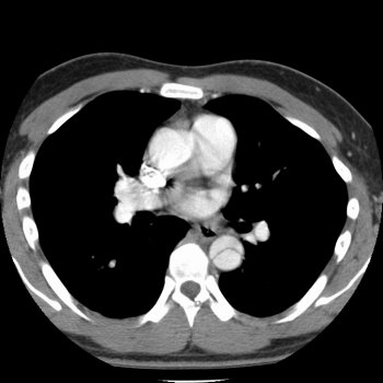
PCP is characterized dyspnea, fever, a nonproductive cough, retrosternal chest pain, tachypnea, tachycardia, few abnormalities on auscultation, bilateral patchy alveolar infiltrates on chest x-ray, and round cysts found under light microscopy when stained with methenamine silver. The treatment is trimethoprim-sulfamethoxazole. The "episode of fever, headaches, joint pain, a loss of appetite, and mild sore throat" that she describes having a few months earlier is consistent with the acute HIV syndrome which affects many patients with HIV, approximately three to six weeks after the primary infection. This syndrome coincides with plasma viremia (wide dissemination of the virus). The symptoms gradually subside over a few weeks.

"Did you or your late husband ever install insulation or brake lining, do construction work, or work in a shipyard?" (choice A) is an important question if an asbestos-related disease is suspected, but this case is more consistent with Pneumocystis carinii pneumonia than asbestosis or malignant mesothelioma. Asbestosis is characterized by dyspnea, a nonproductive cough, basilar crackles or rales, clubbing, linear streaking and pleural thickening seen on chest x-ray, and ferruginous bodies seen on microscopic examination of lung tissue (rod-shaped bodies with clubbed ends). Malignant mesothelioma is a tumor of the pleura that is characterized by chest pain, dyspnea, a cough, a chest x-ray showing pleural fluid, irregular pleural thickening, and a biopsy demonstrating the malignant cells.

"Have you ever been involved in a homosexual relationship?" (choice C) is not a vital question at this time because it is very unlikely that this woman contracted HIV and AIDS from unprotected sexual intercourse with another woman. It is more likely that she contracted the infection from unprotected sexual intercourse with a man. Also, if you wanted to know if a patient has homosexual relationships, it is better to ask in a nonjudgmental way, such as, "Do you have sex with men, women, or both?"

"Have you ever had drink in the morning to get started (an "Eyeopener")?" (choice D) is a part of a four question screening test to detect problem drinking called the CAGE questionnaire. The other three questions that make up the CAGE questionnaire include: “Have you ever felt the need to Cut down on your drinking?”, “Have you ever felt Annoyed by criticisms of your drinking?”, and “Have you ever had Guilty feelings about drinking?”. Two positive responses indicate that a problem is likely. These questions may be important, but the immediate concern in this case is this patient's Pneumocystis carinii pneumonia and whether she has been infected with the HIV virus. Questions related to her sexual practices may help to identify the source of infection.

"Have you ever had a positive PPD or been exposed to anyone with tuberculosis?" (choice E) is a question that is important if tuberculosis or an aspergilloma is suspected. However, this case is more consistent with Pneumocystis carinii pneumonia. Primary tuberculosis (TB) is characterized by systemic symptoms, a cough, sputum production, hemoptysis, lower lobe infiltrates, hilar node enlargement, pleural involvement, and the presence of acid-fast bacilli. Reactivation TB is characterized by infiltrates with cavitation in the apices. An aspergilloma is a "fungus ball"' that typically forms within a preexisting cavity (from TB or sarcoidosis) in the pulmonary parenchyma. The patients may be asymptomatic or present with hemoptysis. A culture shows fungal mycelia, which appear as branching hyphae. In this case, microscopic evaluation shows round structures, not acid-fast bacilli or hyphae.


A 45-year-old man is brought to the emergency department after fainting while walking his dog. He denies a history of chest pain or prior syncope. His past medical history is significant for a cardiac murmur known since childhood. He does not smoke, and denies any recreational drug use. His temperature is 37 C (98.6 F), blood pressure is 128/78 mm Hg, pulse is 87 /min and regular, and respiratory rate is 18/min. He has a prominent apical impulse and a II/VI, late peaking murmur at the right upper sternal border that decreases in intensity with a Valsalva maneuver. An electrocardiogram shows large S waves in leads V1 and V2, and large R waves in leads V4 through V6, with ST depressions in leads V5 and V6. The most likely diagnosis is

  A. aortic stenosis
  B. idiopathic hypertrophic subaortic stenosis (hypertrophic cardiomyopathy)
  C. pericarditis
  D. pulmonary embolism
  E. pulmonary hypertension
Explanation:

The correct answer is A. The presence of a systolic murmur over the aortic area that decreases as preload is decreased (i.e., with a Valsalva maneuver) is consistent with aortic stenosis. The fact that the murmur is late peaking suggests that the stenosis is significant. The prominent apical impulse and EKG suggests compensatory left ventricular hypertrophy (LVH). The ST changes in leads V5 and 6 are often seen with left ventricular hypertrophy from systolic overload, and are termed a “strain pattern.” The presence of syncope with aortic stenosis significantly impacts the mortality associated with this disease. Data suggests that the average time to death from aortic stenosis with syncope is 3 years.

Idiopathic hypertrophic subaortic stenosis (choice B) is unlikely since the murmur decreases with decreasing preload (the Valsalva maneuver). The outflow obstruction associated with idiopathic hypertrophic subaortic stenosis increases with decreasing preload and therefore, a decrease in preload is associated with an increase in the murmur intensity.

Pericarditis (choice C) is unlikely given the absence of chest pain, a precordial rub, or typical EKG findings (diffuse ST elevation, diffuse PR depression with PR elevation in aVL).

Pulmonary embolism (choice D) is unlikely given that the patient has no known history of risk factors for this process (e.g., hypercoagulability, poor activity), is not tachycardic or tachypneic, and has no EKG findings consistent with a pulmonary embolus (sinus tachycardia, S wave in lead I, Q wave and T wave inversion in lead III, or ST-T wave changes in leads V1 through V4).

Pulmonary hypertension (choice E) is unlikely given that there is no history of progressive dyspnea. The physical exam also does not show evidence of pulmonary hypertension (pulmonic tap, prominent pulmonic component of the second heart sound) or right heart strain (right ventricular heave, right sided S4, tricuspid regurgitation, etc).


A 77-year-old woman comes to the emergency department because of a "real bad left-sided headache." She says that she is generally healthy, but over the past few weeks she has experienced fever, fatigue, transient visual loss in the left eye, and scalp pain. Her temperature is 38.2 C (100.8 F), blood pressure is 130/90 mm Hg, pulse is 68/min, and respirations are 18/min. Physical examination shows tenderness over the right temple. The remainder of the examination is unremarkable. The most appropriate next step is to

  A. determine the erythrocyte sedimentation rate
  B. determine Hepatitis B serology
  C. obtain a surgery consult for a renal biopsy
  D. obtain a surgery consult for a temporal artery biopsy
  E. order a CT scan of the head
  F. order an MRI of the head
  G. start immediate treatment with methylprednisolone, intravenously
Explanation:

The correct answer is G. This patient most likely has temporal arteritis (TA), and requires immediate treatment to prevent blindness. TA often affects elderly women and presents with fever, anemia, an elevated erythrocyte sedimentation rate, and a headache. A biopsy typically shows panarteritis with inflammatory mononuclear cell infiltrates and giant cells within the wall of the temporal artery. A segmental biopsy is often necessary to establish the diagnosis, because the disease may be segmental. Serious complications are ocular involvement and blindness, which may be avoided with early corticosteroid therapy. If temporal arteritis is suspected, corticosteroids should be given immediately, even before the biopsy is performed, to prevent blindness. Temporal arteritis is often associated with polymyalgia rheumatica, which is characterized by stiffness, aching, and pain in large muscle groups.

The erythrocyte sedimentation rate (choice A) is often elevated in patients with temporal arteritis (TA), however, this patient has had transient visual loss, which may progress to permanent blindness, and requires immediate treatment even before the diagnosis is established. The work-up can be done after treatment is initiated.

Hepatitis B serology (choice B) is part of the diagnostic evaluation of patients with polyarteritis nodosa (PAN), not temporal arteritis. Patients with polyarteritis nodosa, which is a multi-system necrotizing vasculitis, often have a positive test for hepatitis B surface antigen. These patients often present with fever, weight loss, malaise, and other symptoms that are specific to the involved organs. These patients may have positive p-ANCA titers. A tender temporal artery and transient visual loss are not typically found in polyarteritis nodosa.

A renal biopsy (choice C) is sometimes used to establish the diagnosis of polyarteritis nodosa (PAN), not temporal arteritis. PAN is a multi-system necrotizing vasculitis that often involves the renal arteries. Hypertension is present when the renal arteries are affected. The typical renal finding is arteritis without glomerulonephritis. Treatment is prednisone and cyclophosphamide.

A temporal artery biopsy (choice D) is performed to establish the diagnosis of temporal arteritis. This patient has symptoms that are consistent with this disease and therefore treatment must be started immediately to prevent blindness. A biopsy should be obtained after the corticosteroids are given.

A CT scan (choice E) and an MRI of the head (choice F) are not part of the work-up for temporal arteritis. Temporal arteritis is an inflammatory disorder of medium and large-sized arteries that typically involves the branches of the carotid artery. The diagnosis is confirmed with a temporal artery biopsy. If a tumor, a hemorrhage, cerebrovascular accident, or a transient ischemic attack are suspected, these tests should be considered.


A 76-year-old woman is brought to the hospital by her son because of "rapid breathing." She has advanced Alzheimer disease and is unable to give a coherent history. She was recently diagnosed with breast cancer. She lives alone, but normally has a health care aide during the day. The aide was not available when the son tried to reach her to ask if anything happened. The son has not seen his mother in 2 months. An accentuated fall in systolic blood pressure during inspiration would most likely suggest

  A. anxiety
  B. cardiac tamponade
  C. myocardial infarction
  D. senile aortic stenosis
  E. sepsis
Explanation:

The correct answer is B. This question describes pulsus paradoxus, which is when there is an accentuated fall in systolic blood pressure (>10 mm Hg) during inspiration. There is usually a small decrease in blood pressure during inspiration, but it is exaggerated in cardiac tamponade because the external compression caused by fluid accumulation around the heart leads to impaired ventricular filling, reduced left ventricular stroke volume, and a reduction in systolic blood pressure. Cardiac tamponade can occur acutely after trauma or develop chronically from the accumulation of pericardial fluid malignancies (she has breast cancer), uremia, infections, collagen vascular diseases, or radiation.

All of the other choices, anxiety (choice A), myocardial infarction (choice C), senile aortic stenosis (choice D), and sepsis (choice E), may be associated with tachypnea, but are not usually direct causes of pulsus paradoxus.


A 35-year-old prisoner was recently stabbed in the left leg by another inmate. He is brought to the emergency department by the county corrections officer because of high fevers, swelling of the left thigh, and severe pain at the puncture wound site. His temperature is 38.3 C (101.0 F), blood pressure is 90/50 mmHg, pulse is 120/min, and respirations are 25/min. Although he is awake, he appears lethargic. The left thigh appears pale and swollen around the puncture sight. There is notable crepitus on palpation around the wound. X-rays of the left thigh show translucences in a feathery pattern along the quadriceps. Laboratory studies show:

On exploration of the wound, serosanguinous discharge is noted. Blood cultures are drawn and intravenous crystalloid fluids are instituted. A tetanus booster shot is administered. The most appropriate additional therapy is

  A. ceftriaxone and aztreonam
  B. ciprofloxacin
  C. penicillin and clindamycin
  D. ribavirin and interferon alpha
  E. vancomycin and gentamicin
Explanation:

The correct answer is C. This patient most likely has gas gangrene and myonecrosis. The causative organism is the Gram-positive anaerobic cocci, Clostridium perfringens. The x-ray shows classic feathery gas pattern seen in gas gangrene. The triad of management aside from fluid replacement includes antibiotics, surgical debridement, and hyperbaric oxygen. Penicillin and clindamycin are the most active antibiotic regimen against the Clostridia species.

Ceftriaxone (choice A) is an alternative antibiotic choice to penicillin and clindamycin, but aztreonam lacks any significant activity against Gram-positive organisms.

Ciprofloxacin (choice B) and other fluoroquinolones (except levofloxacin) generally have weak activity against the Clostridium species.

Ribavirin and interferon alpha (choice D) are anti-viral agents, not antibacterial agents.

Vancomycin (choice E) has sufficient coverage for C. perfringens. Gentamicin is an aminoglycoside that requires oxygen-dependent transport into bacterial cells, and so it is ineffective against anaerobic organisms.


A 27-year-old woman comes to the emergency department with "red urine" and a 7-hour history of severe right-sided flank and groin pain. She has no other past medical history and takes only oral contraceptive pills for medications. She had one episode of vomiting in that period of time. She appears otherwise healthy but in moderate discomfort. Her temperature is 37.0 C (98.6 F), blood pressure is 123/90 mm Hg, and pulse is 100/min. Her physical examinaiton is notable for mild suprapubic tenderness but no costovertebral angle tenderness. Her urine is dipstick positive for red blood cells. The most appropriate next step in the evaluation is a/an

  A. abdominal CT scan with contrast
  B. abdominal radiograph
  C. abdominal ultrasound
  D. laparoscopy
  E. stone-protocol abdominal CT
Explanation:

The correct answer is B. This patient clearly has nephrolithiasis. Typical presentations include hematuria, flank pain radiating to the groin, and a benign abdominal exam. Since most kidney stones are composed of calcium oxalate, they are radio-opaque and thus visible on plain radiographs. The remaining components of the diagnostic evaluation include analysis of the urine for crystals, blood, and pH.

An abdominal CT scan with contrast (choice A) is considered not sensitive enough to detect renal stones. However, in some centers, a stone-protocol abdominal CTs (choice E) are being increasingly utilized. The resolution of new CT scanners is such that when properly focused on the appropriate anatomy in high-resolution cuts (1.5mm vs. standard 5 or 10mm cuts), most stones, opaque or lucent can be visualized. However, plain radiographs still remain the first imaging procedure of choice.

An abdominal ultrasound (choice C) has no utility in the diagnosis of renal stones but is very useful in the diagnosis of gallstones.

A laparoscopy (choice D) provides visualization of the abdominal cavity and therefore is of no use in the diagnosis of intrarenal system stones.


A 47-year-old male is brought to the emergency department after he is injured in a fist fight. He was punched in the face multiple times and has pain and swelling around his left eye. Physical examination demonstrates ecchymosis and swelling of his left lower eyelid. There is a mild left periorbital swelling but no obvious tenderness or step off deformity on palpation. The cornea, lens, and anterior chamber are clear bilaterally. The pupils are equal and reactive. There is a mild restriction of upward gaze in his left eye, but there is normal abduction and adduction. Extraocular movements are normal in the right eye. Sensation in the distribution of the infraorbital nerve is intact. A coronal CT scan of the orbits is shown . The most likely complication of this type of orbital injury is

  A. blindness
  B. entrapment of the inferior rectus muscle
  C. entrapment of the medial rectus muscle
  D. an orbital pseudotumor
  E. sinusitis
Explanation:

The correct answer is B. The coronal CT scan of the orbits clearly demonstrates an orbital floor fracture. The patient's exam finding of restricted upward gaze also suggests injury to the inferior rectus muscle. Orbital wall fractures most often occur in the orbital floor and sometimes in the medial wall of the orbit, because these are the weakest regions of the bony orbit. The proximity of the paranasal sinuses, nerves, vessels, extraocular muscles, globe, and other orbital structures predispose them to a wide variety of possible damage from injuries producing orbital fractures. The most common extraocular muscle entrapped in orbital floor fractures is the inferior rectus muscle, which limits upward gaze.

Blindness (choice A) is not a complication of orbital floor fractures. Blindness can occur if there is direct injury to the globe itself or if there is injury to the optic nerve. There are no signs of globe or optic nerve injury in this patient's physical examination or in the CT scan.

Entrapment of the medial rectus muscle (choice C) is a complication of fractures of the medial orbital wall. Entrapment of this extraocular muscle is manifested by limited abduction of the eye.

An orbital pseudotumor (choice D) is not associated with orbital injury. It is an idiopathic granulomatous inflammation of the orbit that often presents with chemosis, proptosis, and pain. The most common structure in the orbit to be involved is the lacrimal duct.

Sinusitis (choice E) is not associated with orbital injury. Sinusitis is often due to underlying allergies. Orbital floor fractures or other fractures of the orbit do not predispose patients to the development of sinus disease.


A 28-year-old man comes to the emergency department with a 2-day history of worsening abdominal pain and a lack of desire to eat. He was diagnosed as having leukemia 4 months ago, for which he received chemotherapy successfully. For the past 2 months he has been asymptomatic and able to tolerate a regular diet. In the emergency department, he is noted to pass bloody mucoid stools and hematuria. His temperature is 37.8 C (100 F), blood pressure is 120/70 mm Hg, and pulse is 92/min. Abdominal examination shows a diffusely tender abdomen with voluntary guarding and no peritoneal signs. Laboratory studies show a total leukocyte count of 500/mm3 and a platelet count of 15,000/mm3. A subsequent CT scan of the abdomen and pelvis reveals thickening of the sigmoid colon with minimal infiltration of the surrounding fat. The most appropriate management of this patient is

  A. antibiotics
  B. antibiotics, bowel rest, platelet transfusion, and granulocyte stimulating factor
  C. antibiotics, colonoscopy
  D. colonoscopy
  E. granulocyte stimulating factor, antibiotics, and colonoscopy
Explanation:

The correct answer is B. This patient was recently treated with chemotherapy for leukemia and has developed a neutropenic enterocolitis. Neutropenic enterocolitis is usually segmental, often localized to the sigmoid colon. This can be diagnosed on a CT scan of the abdomen and pelvis by means of a thickened sigmoid colon and infiltration of the surrounding fat. Without being treated with antibiotics and bowel rest, this neutropenic enterocolitis can progress to worsening of the symptoms and signs leading to complications of systemic septicemia and hypotension. Hence, this patient should be treated with bowel rest, appropriate antibiotics, and platelet transfusion to increase his platelet counts and decrease the chance of hematuria. Granulocyte stimulating factor may help to increase his neutrophil count.

Antibiotics (choice A) alone may not resolve colitis completely without the correction of neutropenia.

Colonoscopy (choices C and D) is contraindicated as the patient may develop complications from colonoscopy of an inflamed colon.

Granulocyte stimulating factor (choice E) is indicated to increase the neutrophil count but colonoscopy is contraindicated.


A 27-year-old man is brought to the emergency department after being stabbed in the chest with a knife at a local bar. He suffered multiple kicks to the abdomen and a stab wound with an unknown type of blade to the right chest. The patient's past medical history and allergies are unknown. The patient last ate 3 hours ago. He has large patches of dried blood on his shirt, face and lips. He is diaphoretic but speaking in full sentences. He has multiple stab wounds on his right chest both inferior and superior to the right nipple. The most appropriate first step in management is to

  A. assess the patient for additional cardiac sounds
  B. assess the patient's airway
  C. assess the patient's blood pressure
  D. assess the patient's jugular venous pulsations
  E. assess the severity of the stab wound
Explanation:

The correct answer is C. In trauma situations, the most important skill is to keep focused on the ABC and D's of patient evaluation. There is an overwhelmingly strong tendency to pay attention to the obvious and forget to further evaluate a patient. This is the most assured way to inflict further harm on a patient. Assessment of this patient's blood pressure is appropriate at this time.

Assessing the patient for additional cardiac sounds (choice A) can be done later on, however at this time it is most important to evaluate blood pressure.

In this case, since the patient is speaking in full sentences, evaluation of his airway (choice B) is not relevant and it is critical to assess his blood pressure.

Assessment of the patient's jugular venous pulsations (choice D) is a more advanced physical examination maneuver that may be performed once the initial trauma survey is completed. It is not performed during the initial survey.

Exploration of the region of the stab wound (choice E) will be done surgically in the operating room. There is no place for this examination during the phase of patient care devoted to primary survey and assessment.


One winter evening you are evaluating a 68-year-old woman who is complaining of shortness of breath in the emergency department. She has a medical history significant for chronic obstructive pulmonary disease (COPD) and hypertension. Her medications include an ipratropium and albuterol inhaler and furosemide. Following a series of albuterol nebulizer treatments, her respiratory function returns to baseline. Her temperature is 37.0 C (98.6 F), blood pressure is 146/87 mm Hg, pulse is 89/min, and respirations are 22/min. She has diminished breath sounds bilaterally, but otherwise her lungs are clear to auscultation. Prior to her discharge, she should receive

  A. a tetanus booster
  B. vaccination against hepatitis B
  C. vaccination against influenza
  D. vaccination against Haemophilus influenzae
  E. vaccination against varicella
Explanation:

The correct answer is C. Elderly patients with underlying lung disease are at high risk for severe influenza infections during the wintertime. Therefore, this patient population must receive their influenza vaccine in order to reduce their risk.

A tetanus booster (choice A) is not warranted since this patient has no history of trauma and exposure to contaminated elements (soil etc.)

Vaccination against hepatitis B (choice B) is not warranted in this instance since the patient has no history of exposure to possible contaminated products such a blood or bodily fluids.

Vaccination against Haemophilus influenzae (choice D) is typically reserved for children. It is not routinely given to adults.

Vaccination against varicella (choice E) is not warranted since there is no history of exposure to the varicella zoster virus during the current admission or recent past.


An 11-year-old boy is rushed into the emergency department following a motor vehicle accident. The patient was a restrained, front seat passenger when an oncoming car hit the passenger side of the car. The boy denies loss of consciousness, but in the hospital he complains of pain over his right side. His pulse is 139/min, blood pressure is 118/59 mm Hg, and respirations are 24/min. On physical examination he has decreased breath sounds over the right base and there is ecchymosis over the right flank. His abdomen is soft with tenderness in the right upper quadrant. Appropriate management of his airway with neck stabilization is provided and he is resuscitated appropriately. Imaging studies of his neck are negative and a chest X-ray does not show a pneumothorax or rib fracture. Dipstick of spontaneously voided urine is positive for blood. Urinalysis confirms the presence of 50 RBCs/hpf. In regards to his hematuria the next most appropriate course of action is to

  A. begin empiric antibiotics
  B. obtain CT scan of abdomen and pelvis
  C. order renal/bladder ultrasound
  D. perform retrograde urethrogram
  E. place Foley catheter
  F. repeat urinalysis
  G. schedule outpatient intravenous pyelogram (IVP)
Explanation:

The correct answer is B. The management of hematuria associated with trauma differs in adults and children. In the adult population, imaging is performed only in those patients with gross hematuria or microscopic hematuria plus hypotension. This differs from the pediatric patient. In children, any degree of hematuria (gross or microscopic) should be investigated with imaging studies. One reason for this discrepancy is that large amounts of catecholamines released in injured children may sustain blood pressure in the face of hypovolemia. A CT scan is the most useful imaging modality in this setting. A CT is noninvasive, accurate and fast, and it can help in assessing the size and extent of retroperitoneal hematomas and renal parenchymal trauma. Not only does this child have microscopic hematuria (an indication by itself to perform imaging studies), but he also has signs (flank ecchymosis and tenderness) that raise the suspicion of renal injury. High suspicion for renal injury (i.e., rib fracture, flank contusion, deceleration injury) is another indication to perform imaging studies.

There is no indication at this time that the patient requires antibiotics (choice A). He has no open fractures, large abrasions, or burns. Further diagnostic studies must be undertaken to determine if the patient requires antibiotics for any disruption to the urinary system.

Ordering a renal/bladder ultrasound (choice C) acknowledges the fact that the patient does require investigation of his urinary system. However, a CT scan is quicker and will have a much higher yield for associated urinary and abdominal injuries versus ultrasound.

A retrograde urethrogram (choice D) allows for visualization of the urethra to investigate for extravasation. It is performed when there is gross blood at the meatus. It will not help in this patient whose injuries are suspected to be intraabdominal.

Hematuria is not an indication for Foley catheter placement (choice E). As long as the patient is awake without altered sensorium he should be given an opportunity to void on his own (which he has done). It should be noted that in any patient who has gross blood at the meatus a retrograde urethrogram must be obtained prior to placing a Foley catheter.

Repeating the urinalysis (choice F) will only delay this patient's work-up. Although the urine dipstick may be falsely positive for heme, there is no reason to doubt the validity of the urinalysis. Besides, this patient's flank ecchymosis and tenderness warrant imaging independently of his urinalysis results.

Intravenous pyelogram, or IVP (choice G), has a role in evaluating the urinary tract. However, with the use of CT scan, IVP has a very limited role in evaluating renal/ureteral trauma. Delaying imaging until this patient is an outpatient could mean missing a potentially life-threatening renal injury.


A previously healthy 3-week-old baby is brought by his parents to your emergency department with a 1-day history of emesis. The parents describe the emesis as "forceful", non-bloody, and non-bilious. The baby is exclusively breastfed and continues to be hungry after each episode of vomiting. They deny any fevers. You notice an active baby boy with unremarkable vital signs. Physical examination is significant for a peristaltic wave on the abdomen and a 2x2 cm firm mass palpated in the midepigastric region. Laboratory studies show a bicarbonate level of 18 mEq/L. The most likely diagnosis is

  A. annular pancreas
  B. gastroesophageal reflux
  C. intussusception
  D. malrotation with volvulus
  E. pyloric stenosis
Explanation:

The correct answer is E. This patient presents with a classic case of pyloric stenosis. It is the most common surgical condition seen in the newborn period. It is more common in males than females. It is caused by a hypertrophic pyloric muscle, which causes obstruction of the gastric outlet. This hypertrophic muscle can be palpated in the midepigastric region and represents the classic "olive" associated with pyloric stenosis. The emesis is non-bilious because the obstruction is proximal to the ligament of Treitz. Once the diagnosis is made, treatment consists of a pyloromyotomy.

Annular pancreas (choice A) is caused by the failure of the resorption of the left pancreatic bud during rotation of the duodenum during fetal development, which causes a ring-like structure around the duodenum, often the second portion. These patients present with signs of partial or complete small bowel obstruction and will have frequent bilious vomiting and abdominal distension often in the first week of life. There is often a history of polyhydramnios.

Patients with gastroesophageal reflux (choice B) rarely present with projectile vomiting. Parents often complain of spitting up with feeds, arching of the back, and waking up from sleep crying. Mild GER often responds to changing to an upright feeding position and thickening the feeds with cereal. Moderate GER may require medication, such as an H2-blocker, for control. Severe reflux may require surgical intervention.

Patients with intussusception (choice C), caused by an invagination of the bowel, classically present with colicky abdominal pain, raising of their knees to their chest to relieve this pain, a sausage-like abdominal mass, and "current jelly" stools. They may also have vomiting that begins as non-bilious and progresses to bilious as the obstruction progresses.

Malrotation with volvulus (choice D) is caused by failure of the small intestine and right colon to reach their proper anatomic location during fetal development and twisting of the gut, resulting in compromised perfusion to this area. These patients have bilious vomiting, may have bloody stools, and appear quite ill as compromised perfusion progresses to infarction and gangrene.


A 64-year-old woman comes to the emergency department with a 36-hour history of diffuse abdominal pain, abdominal fullness, nausea, and vomiting. She has no appetite and is unable to eat or drink secondary to nausea and vomiting, which is bilious in color. She passed loose brown stool earlier today. She denies any bright red blood per rectum or bloody vomitus. Her past medical history is notable for endometrial cancer 4 years ago treated with surgery and radiation. The patient denies ever experiencing similar symptoms in the past. Her temperature is 37.0 C (98.6 F), blood pressure is 110/70 mm Hg, pulse is 100/min, and respirations are 16/min. She has a moderately distended abdomen with diffuse tenderness on palpation. There is no rebound tenderness or guarding. Bowel sounds are high-pitched. There is no occult blood on rectum examination. Initial laboratory studies show:

The next most appropriate step to confirm the diagnosis is to obtain

  A. a CT scan of the abdomen
  B. plain films (supine abdomen x-ray and upright chest and abdomen x-ray)
  C. a right upper quadrant ultrasound
  D. serum amylase and lipase
  E. serum lactate
Explanation:

The correct answer is B. This patient most likely has a small bowel obstruction as evidenced by prior abdominal surgery, a diffusely tender abdomen, high-pitched bowel sounds, and nausea and vomiting. Small bowel obstruction usually results from either mechanical blockage or paralytic ileus. Postoperative adhesions or an incarcerated inguinal hernia are the most common causes of a mechanical small bowel obstruction. This patient is at risk for bowel obstruction given her prior abdominal surgery and radiation, thus putting her at increased risk for adhesion and stricture formation. Patients with small bowel obstruction typically present with a distended, tender abdomen, nausea and vomiting, and high-pitched bowel sounds. The presence of peritoneal signs suggests infarcted bowel or perforation. Laboratory tests may reveal a normal or elevated white blood cell count, elevated hematocrit secondary to hemoconcentration, and low potassium and chloride secondary to vomiting. Blood urea nitrogen and creatinine may be elevated secondary to prerenal azotemia. Plain films are very helpful in diagnosing small bowel obstruction and should be part of the initial management of the patient. X-rays of the abdomen commonly reveal gas-filled, distended loops of bowel with air-fluid levels. An upright chest x-ray is needed to evaluate for possible bowel perforation which is demonstrated by the presence of free air under the diaphragm.

A CT scan of the abdomen (choice A) is rarely needed to diagnose a small bowel obstruction and can worsen a patient's symptoms, especially in the setting of a complete small bowel obstruction. In complete obstruction, administering contrast from above will add to the degree of fluid and edema proximal to the obstructed bowel. A CT scan of the abdomen may be helpful later in a patient's course. For example, it may be necessary to obtain a CT scan to exclude the possibility of a tumor compressing bowel that leads to obstruction. This imaging modality, however, is not part of the immediate management.

A right upper quadrant ultrasound (choice C) is used to image the liver, gallbladder, and ducts. The patient does not have any abdominal symptoms or pain localized to this region to warrant starting with this imaging modality. An uncommon but important cause of small bowel obstruction in elderly patients is gallstone ileus. In this setting, a gallstone erodes through the gallbladder wall and into the small bowel, causing intraluminal obstruction. Typically, patients will give a history of right-sided upper abdominal pain at the onset of their symptoms and may demonstrate air in the biliary tree on ultrasound. This patient, however, does not have any symptoms to suggest the presence of gallstones.

An elevated serum amylase and lipase (choice D) is useful in diagnosing pancreatitis. Pancreatitis usually presents with epigastric pain radiating to the back, as well as nausea and vomiting. The two leading causes of pancreatitis are alcohol abuse and gallstones. This patient's presentation and exam are not classic pancreatitis as described above.

An elevated lactate (choice E) is frequently elevated in the setting of hemorrhage, shock, sepsis, and hypoxia. This patient has an elevated anion gap of which suggests the presence of a lactic acidosis. An elevated lactate, however, may suggest many processes as outlined above and thus would not be helpful in confirming the diagnosis of small bowel obstruction.


You are called to the emergency department to evaluate a 6-year-old girl who has developed a rash on her distal extremities 2 days ago that has been progressing toward her trunk. She has had a fever and arthralgias over the past 2 days for which she was given acetaminophen. On further questioning, the patient's mother reports that the patient was bitten by her pet rat a few days prior to onset of the fever and rash; however, the site appears to be healing well. Her mother reports that the girl has a normal past medical history without any significant health problems. Laboratory studies show leukocytosis with an elevated neutrophil count. Blood culture results are pending. At this time the most correct statement about this patient's condition is:

  A. Her rash is classic for Rocky Mountain Spotted Fever and she should be started on antibiotics for rickettsial organisms
  B. Meningitis can be ruled out because the rash usually starts on the trunk and extends peripherally
  C. The patient should be admitted and started on broad-spectrum antibiotics to cover Streptobacillus moniliformis and Spirillum minor
  D. This is a drug eruption secondary to acetaminophen and you should advise the patient's mother to stop acetaminophen and give ibuprofen instead
  E. This is most likely a viral exanthem and will resolve spontaneously in 1-2 weeks
Explanation:

The correct answer is C. Rat bite fever is an acute febrile illness that is usually accompanied by a skin rash. It is caused by either Streptobacillus moniliformis or Spirillum minor which are the bacteria that generally infect humans as a result of the bite of a rat, mouse, or other rodents. Following a rat bite, there is minimal local inflammation with prompt healing of the wound. If the infection is not halted, bacteremia ensues and lesions distant from the bite appear 1-3 days after the bite. Manifestations include fever, rigors, headaches, malaise, and arthritis. Treatment of choice is penicillin G or tetracycline for patients allergic to penicillins. Occasionally the course is complicated by endocarditis, meningitis, myositis, abscesses, splenic or renal infarction, brain abscess, and sepsis.

Rocky mountain spotted fever (choice A) is incorrect because there is no history of tick bite reported. In addition, in a child of this age group, fever accompanied by an extremity rash should alert physicians to treat and cover meningitis organisms until blood culture results become available. Therefore, since meningitis cannot be definitively ruled out at this time, (choice B) is also incorrect.

Drug eruption (choice D) is incorrect for two reasons. First, drug eruptions typically start on the trunk and extend peripherally. Second, fever and arthritis usually do not accompany typical drug eruptions.

Viral exanthem (choice E) is incorrect at this stage of evaluation. One should rule out more urgent causes of fever and rash before concluding that it is viral exanthem.


A 45-year-old man is struck by a motor vehicle and is transported by a Med-Flight to the local emergency department. The patient is reported to be intoxicated with a Glasgow score of 15 on the scene of the accident. He was struck by a vehicle while crossing the street. On arrival, the patient is awake and somewhat combative. He is alert to person only. Primary survey reveals a well-developed man in mild distress. He is in a cervical collar. His blood pressure is 150/90 mm Hg, his heart rate is 130 /min, and he is breathing at 26/min. He has obvious lower extremity tibia fractures bilaterally and a laceration on his forehead. Secondary survey reveals severe pelvic trauma with bilateral inferior and superior pubic ramus fractures. The most appropriate diagnostic test at this time is

  A. abdominal CT scan
  B. abdominal ultrasound
  C. diagnostic peritoneal lavage
  D. exploratory laparotomy
  E. pelvic ultrasound
Explanation:

The correct answer is A. This patient has severe trauma involving his lower extremities and pelvis. The concern is for intraabdominal trauma resulting in bleeding. An abdominal CT scan is the imaging modality of choice since it detects both intraabdominal and retroperitoneal blood.

An abdominal ultrasound (choice B) has been shown to be equivalent to diagnostic peritoneal lavage (choice C) for detection of free intraabdominal blood. However, neither of these modalities can reliably diagnose the presence of a retroperitoneal bleed.

Although some centers immediately opt for an exploratory laparotomy (choice D), this is actually not the appropriate diagnostic step. This patient will certainly go to the operating room for repair of his tibial fractures and stabilization of his pelvis. The addition of a midline abdominal incision to his procedure list would entail a tremendous amount of OR time and additional chance of morbidity for the patient. The most appropriate intervention is to look for the presence of blood and if the clinical picture still suggests a bleed, but there has been none disclosed by imaging, then a surgical exploration is warranted.

A pelvic ultrasound (choice E) offers no diagnostic benefit for the evaluation of an intraabdominal bleed.


A 27-year-old man comes to the emergency department because of increasing fatigue, malaise, chills, and low-grade fevers over the last 2 weeks. He reports no recent sick contacts and denies any significant past medical history. The patient does mention that he uses heroin frequently but not since last week. His temperature is 38.8 C (101.8 F), blood pressure is 85/60 mm Hg, and heart rate is 120/min. On physical examination, the patient appears gaunt, malnourished, and dehydrated. A faint systolic murmur is audible on cardiac auscultation. Needle tracks are found at both antecubital fossa. Petechiae are noted across his back and splinter hemorrhages are found under the nail beds of his right hand. Laboratory studies show:

A chest radiograph shows normal lungs and cardiac silhouette. An electrocardiogram reveals sinus tachycardia. Urinalysis shows 2+ proteinuria, 3+ red blood cells, and 1+ ketones. The patient is admitted to the hospital where he becomes progressively more confused and disoriented. Three sets of blood cultures are drawn and intravenous fluids are initiated. The most appropriate next step in management is to

  A. begin nafcillin and gentamicin, intravenously
  B. begin penicillin and clindamycin, intravenously
  C. obtain an immediate evaluation by cardiac surgery
  D. order an urgent transesophageal echocardiography
  E. start methadone therapy
Explanation:

The correct answer is A. This case describes a patient with acute bacterial endocarditis that rapidly deteriorates soon after his initial presentation. He presents with vague medical complaints and a history of intravenous drug use, namely heroin. The initial medical symptoms and vital signs can be consistent with opiate withdrawal. However, his physical exam reveals a murmur, truncal petechiae, and splinter hemorrhages that are indicative of severe endocarditis. In addition, he has an elevated white blood cell count. The urinalysis and elevated creatinine indicate possible glomerulonephritis from immune-complex deposition. Last of all, he has a notable change in his mental status. All these data support the diagnosis of acute bacterial endocarditis. Due to his rapidly worsening clinical course, it is imperative to begin empirical antibiotic treatment once blood cultures are drawn, since early antibiotic therapy may prevent further morbidity and sepsis. In intravenous drug users, the most common pathogen is Staphylococcus aureus. Other organisms include Staphylococcus epidermidis, Streptococcus viridans, Enterococcus, and various Gram-negative rods. Since the organism is unknown, broad antibiotic coverage is indicated. Antibiotic therapy with nafcillin and gentamicin is a typical combination for native valve endocarditis. If MRSA is prevalent, vancomycin and gentamicin are an alternative combination.

Penicillin and clindamycin (choice B) are excellent antibiotics for Gram-positive organisms, particularly the Streptococcus species and anaerobic bacteria. However, Staphylococcus aureus is the predominant organism that causes acute bacterial endocarditis in intravenous drug users, and neither antibiotic covers Staphylococcus aureus well.

Cardiac surgery consultation (choice C) to evaluate for valvular replacement surgery is indicated only if there is invasive infection, fungal infection, recurrent systemic emboli, refractory infection, or refractory heart failure present. Further evaluation of the patient will be required to determine if this is necessary.

An echocardiogram (choice D) is required for definitive diagnosis and for further assessment of the valve to rule out paravalvular abscesses or invasive infection. However, this patient is unstable and there is enough clinical evidence to support the diagnosis, so obtaining an echocardiogram will only confirm diagnosis and should not delay treatment with antibiotics. It is usually done after blood cultures are obtained and they are positive.

Methadone therapy (choice E) should be instituted for patients with opiate withdrawal symptoms. However, this patient's acute condition is due to bacterial endocarditis.


A 7-year-old boy is brought to the emergency department by his mother because of "tea-colored urine" for the last several days. He has also had some nausea and vomiting, and his eyes appear swollen when he wakes up in the morning. The eye swelling tends to resolve over the course of the day. He is generally very healthy and there is no family history of any chronic diseases. His temperature is 36.7 C (98.0 F), blood pressure is 130/90 mm Hg, pulse is 96/min, and respiratory rate is 16/min. Physical examination is unremarkable. A urinalysis shows red cell casts. At this time the most appropriate study to confirm your diagnosis is

  A. antinuclear antibody
  B. antistreptolysin O antibody
  C. renal biopsy
  D. renal ultrasound
  E. urine culture
Explanation:

The correct answer is B. Poststreptococcal glomerulonephritis is the most common cause of acute glomerulonephritis in children. It usually follows a streptococcal pharyngitis by 1-2 weeks and a streptococcal skin infection by 2-3 weeks. It most commonly occurs in school-age children and has a male to female predominance of 2:1. It is most commonly characterized by hematuria (microscopic or gross) with red cell casts, proteinuria, hypertension (from fluid overload secondary to decreased glomerular filtration rate), and edema (from retention of salt and water). Laboratory values are usually significant for markedly decreased complement levels (C3 and C4), hypo- or hypernatremia, and a blood urea nitrogen elevated disproportionately to the creatinine. In order to diagnose poststreptococcal glomerulonephritis with certainty, there needs to be evidence of a preceding streptococcal infection such as an elevated ASO or streptozyme.

Systemic lupus erythematosus (SLE) nephritis is another cause of hematuria. If this child had persistently low complement levels, an ANA (choice A) would be indicated as one of the first screening tests for SLE. Keep in mind that only 25% of cases of SLE are diagnosed within the first two decades of life, the male-to-female ratio is 1:8, and renal disease is not present in all cases of SLE. Therefore, it is unlikely that this 7-year-old boy has new onset SLE, and other more common causes of glomerulonephritis must be ruled out first.

At this time, a renal biopsy (choice C) is not indicated. A renal biopsy is an invasive procedure with inherent risks. As this patient fits the perfect description for post streptococcal glomerulonephritis a renal biopsy would only be indicated if the complement level does not return to normal within 8 weeks, and the gross hematuria does not resolve over several weeks. If one was to do a renal biopsy on a patient with post-streptococcal glomerulonephritis, the findings would be: mesangial and capillary cell proliferation, inflammatory cell infiltration, and granular "humps" of IgG and C3 below the glomerular basement membrane.

A renal ultrasound (choice D) is useful in evaluating parenchymal disease, urinary tract abnormalities, or renal blood flow. In the case of post-streptococcal glomerulonephritis, the changes are microscopic and therefore would not be seen on ultrasound.

Urinary tract infections are a common cause of hematuria. Therefore, it is a good idea to do a urine culture (choice E) in all cases of hematuria. Unfortunately, the child in this case has red cell casts which indicate the kidneys are the source for the blood. So, although a urine culture should be sent on this child, it will probably not help with the diagnosis.


A previously healthy 20-year-old man comes to his college medical clinic for headaches and low-grade fevers. He is discharged home with the diagnosis of a "viral syndrome" and instructed to get ample rest. Approximately three hours later his roommate calls 911 reporting that his friend is unconscious and not arousable. On arrival the paramedics find a lethargic, febrile man lying on the floor and unresponsive. The patient is stabilized and he is rushed emergently to the local hospital where an abdominal CT scan shows bilateral adrenal hemorrhages. His blood pressure is 80/40 mm Hg and his pulse is 110/min. He appears very ill and continues to be non-responsive. The most appropriate study at this time is a/an

  A. Cort-Stim test
  B. cortisol level
  C. high-dose ACTH stimulation test
  D. insulin response test
  E. low-dose ACTH stimulation test
Explanation:

The correct answer is B. This patient has bilateral adrenal hemorrhages in the setting of fulminant disseminated Neisseria sepsis. This is called the Waterhouse-Friderichsen Syndrome. The patient therefore almost certainly has acute adrenal insufficiency. In a critically ill patient, a random cortisol level that is low confirms this diagnosis.

A Cort-Stim test (choice A) is a test whereby synthetic ACTH is administered and the cortisol response is measured one hour later. It is more sensitive at detecting adrenal insufficiency but is both too time-consuming and unnecessary in this patient. The threshold for supplying exogenous steroids is very low so all that is needed is a suspicion for insufficiency that a random cortisol level can easily provide.

A high-dose ACTH stimulation test (choice C) or low-dose ACTH stimulation test (choice E) are both very sensitive tests that require 24 hours to perform. They both are useful at quantifying adrenal function so that differentiation between various causes of adrenal insufficiency can be made. However, they are not the most appropriate studies in this acute situation.

The insulin response test (choice D) is another method used to indirectly assess adrenal function but will not be useful in this patient since the goal is rapid direct assessment of her adrenal function.


A 27-year-old man comes to the emergency department 30 minutes after stepping on a nail in his garage. He was running late for his tennis game and he was about to get into his car when he felt a sharp, dagger-like feeling in his left foot. He looked down and saw a 3 inch nail protruding through the sole of his tennis sneaker. He ran back into the house and had his wife drive him to the hospital. He pulled the nail out of his foot in the car and says that the nail appears intact. He says that he is generally very healthy and has not been to the doctor in "ages". He cannot even remember his last "check-up", but he assumes that it was when he was in his "late teens." He does not know his immunization history but he recalls that he had all of the recommended vaccines before going to college. Physical examination shows a clean, stellate puncture wound on his left heel that appears to only penetrate the superficial epidermis. There is no swelling or pain with the movement of the toes and sensation is intact. He shows you the intact, shiny, clean nail. After you irrigate, debride, and carefully inspect the wound, the most appropriate next step is to

  A. advise him to soak his foot in an iodine solution at home
  B. give him a tetanus and diphtheria toxoid
  C. order a CT scan of his left foot
  D. request an immediate orthopedic surgery consult
  E. send him home with a prescription for an antibiotic that covers Pseudomonas aeruginosa
Explanation:

The correct answer is B. This patient has a puncture wound through a sneaker, which is a very common presentation in the hospital and on the boards. The major considerations in this case are immunization history and penetration through the sneaker (risk of Pseudomonas aeruginosa infection). Since he has not been to the doctor in a while, he probably needs a tetanus and diphtheria toxoid at this time. This is recommended over just the single antigen tetanus toxoid for persons >7 years old. The tetanus immune globulin is an antitoxin that is usually only recommended for contaminated, severe wounds, and when the patient is unsure if they have ever received a tetanus vaccination or have an incomplete immunization history. It should be given in this case because it is also recommended for puncture wounds. Since this patient was up-to-date on his immunizations before he went to college, approximately 10 years ago, he requires the Td toxoid. The issue of a Pseudomonas aeruginosa infection is unsure at this time, because he recently stepped on the nail, and there does not seem to be any signs of infection (erythema, warmth, discharge, etc). Careful follow-up is mandatory if he is not started on an antibiotic.

Advising him to soak his foot in an iodine solution at home (choice A) is not the most appropriate next step because this patient stepped on a nail and requires a Td toxoid at this time. Also, some believe that using an iodine solution does not prevent infection and may even delay healing.

A CT scan of his left foot (choice C) is unnecessary at this time. First of all, it appears as if the nail is intact and there is no foreign material left in his foot. But if you were to believe that some metal might be left in the wound, a plain x-ray can be used to show metallic material. A CT scan is used for plastic, wood, and other substances that are radiolucent.

It seems unnecessary to request an immediate orthopedic surgery consult (choice D) for this simple, shallow, clean puncture wound on this patient's heel. If the wound were deep and dirty or if were through the metatarsal head (and the tennis sneaker), orthopedic surgery consultation should be sought. However, this patient still requires a Td toxoid at this time, even if you decide to call for the consult.

Sending him home with a prescription for an antibiotic that covers Pseudomonas aeruginosa (choice E) is inappropriate at this time because he requires a Td toxoid. The question as to whether or not you should prescribe an antibiotic for this clean wound, after administering the toxoid is a tough one. Technically, an antibiotic should be prescribed when signs of infection are present, but since Pseudomonas osteomyelitis can be devastating if it occurs, many people would probably prescribe an antibiotic to try to prevent an infection. In either case, this answer is still incorrect because you cannot send him home with an antibiotic without providing the Td toxoid.


You are working in the emergency department and asked to see an 82-year-old man who was brought in by his wife because of a new, severe headache. His wife tells you that the patient awoke a week ago in the middle of the night with bad left-sided headache and periorbital pain that has persisted ever since. He denies an increase in pain with cough or sneeze or any neck pain. The wife has been giving him acetaminophen, which seems to help a bit. Over the last 2 days he has noticed that his jaw "gets tired" when he is chewing. He denies other neurologic symptoms, including facial and limb numbness or weakness and denies any changes in speech. Examination shows mild temporal tenderness on the left. Laboratory results are significant for a normal cell count and an erythrocyte sedimentation rate of 117 mm/hr. A CT scan of the head is normal. The most appropriate next step in management is to

  A. arrange an immediate temporal artery biopsy
  B. give intravenous prochlorperazine
  C. prescribe a small amount of oxycodone and follow up in clinic in 2 weeks
  D. prescribe a soft cervical collar, diazepam, and follow up in clinic in 2 weeks
  E. prescribe gabapentin and follow up in clinic in 2 weeks
Explanation:

The correct answer is A. This presentation is typical for temporal (giant cell) arteritis. It can cause blindness or stroke if not treated early. The diagnosis can be made by temporal artery biopsy and steroids are typically given to avoid complications.

Intravenous prochlorperazine (choice B) may be used to treat an acute migraine headache. New diagnosis of migraine is rare in an 82-year-old and other causes should be ruled out.

It would be incorrect to merely treat his pain and send him home (choice C). He may be blind in 2 weeks if you miss the diagnosis of temporal arteritis.

Soft cervical collar, diazepam, and follow up in clinic in 2 weeks (choice D) would be the treatment for a muscle contraction headache.

Gabapentin (choice E) may be used to treat chronic pain, it is not the treatment for temporal arteritis.


A 19-year-old woman is brought to the emergency department by her roommate because of confusion. The patient is uncooperative, so the roommate tries to give you as much information as possible. She tells you that she knows that the patient has been smoking pot, drinking alcohol, and "doing some other drugs" since she failed her pre-medical chemistry course last semester. She has been making monthly trips to her grandfather's house lately, taking him to the doctor and then the pharmacy to pick up his medications that are prescribed for various conditions, such as hypertension, gout, insomnia, depression, nasal congestion, and back pain from prostate cancer. Her temperature is 37.0 C (98.6 F), blood pressure is 90/60 mm Hg, pulse is 40/min, and respirations are 6/min. She lapses into coma during the physical examination, making it very difficult to evaluate her, however, you note that she has miosis. The grandfather's medication that is most likely responsible for this patient's condition is

  A. indomethacin
  B. nortriptyline
  C. phenelzine
  D. pseudoephedrine
  E. sustained-release oxycodone
Explanation:

The correct answer is E. This patient most likely has opioid toxicity, which is most likely due to snorting or injecting crushed sustained-release oxycodone pills. The signs of opioid intoxication are miosis, respiratory depression, coma, hypotension, and bradycardia.

Indomethacin (choice A) is a nonsteroidal anti-inflammatory drug that may lead to gastrointestinal bleeding when taken in large doses. The triad of miosis, respiratory depression, and coma is suggestive of opioid intoxication, not NSAID intoxication.

Nortriptyline (choice B) is a tricyclic antidepressant agent that leads to hyperthermia, tachycardia, either hypertension or hypotension, mydriasis, and a prolonged QRS interval on an electrocardiogram. The triad of miosis, respiratory depression, and coma is suggestive of opioid intoxication, not a tricyclic overdose.

Phenelzine (choice C) is an antidepressant monoamine oxidase inhibitor (MAOI) that causes hypertensive crises and the serotonin syndrome (hypertension, tachycardia, fever, coma, and possibly death) when combined with tyramine-containing food (cheese) and serotonin-altering drugs. Miosis and hypotension are not typically seen with an overdose of phenelzine.

Pseudoephedrine (choice D) is a decongestant that may lead to mydriasis, tachycardia, hypertension, hyperthermia, and seizures if taken in increased doses.


A 40-year-old man with hypertension comes to the emergency department complaining of a severe headache that started the previous evening while exercising at the gym. He states that the headache came on suddenly and describes a constant throbbing pain on the top of his head and down the back of his neck. He did not experience any loss of consciousness, but does complain of nausea and says that he vomited twice last night. He also complains of blurry vision, dizziness, and fatigue. He experienced a similar, but less severe headache one week ago that resolved spontaneously after about 2 hours. He has tried ibuprofen and some acetaminophen with codeine that he had left over from a previous tooth extraction, but has not had any relief of his symptoms. He waited to come to the hospital this morning because he thought the headache was due to the stress he has been experiencing at work and that it would resolve on its own. He smokes 1 pack of cigarettes per day, drinks alcohol occasionally, and does not do any illicit drugs. His temperature is 37.0 C (98.6 F), blood pressure is 158/90 mm Hg, pulse is 62/min, respirations are 18/min, and oxygen saturation is 99%. Physical examination shows a well-developed man lying on the stretcher with his right hand covering his eyes in obvious distress secondary to pain. His pupils are equal, round, and reactive to light and accommodation however, you are unable to examine of the fundi due to the patient's discomfort with the light. Cranial nerves II-XII are intact. He has some neck stiffness with flexion and extension. The remainder of the physical and neurologic examination shows difficulty with bilateral finger to nose precision as well as heel to shin coordination. You order a non-contrast CT scan of his head, which is read as normal. The most appropriate next step in the management of this patient is to

  A. administer intravenous analgesia with morphine and metoclopramide and observe in the emergency department
  B. administer subcutaneous sumatriptan and discharge home to follow up with a neurologist
  C. obtain an urgent neurosurgical consultation and a cerebral angiogram
  D. perform a lumbar puncture with CSF analysis
  E. request psychiatric evaluation for acute stress reaction
Explanation:

The correct answer is D. In this patient, you should perform lumbar puncture with CSF analysis for suspected subarachnoid hemorrhage (SAH). The CT scan can be normal in 5% of patients with a subarachnoid hemorrhage within the first 12 hours of headache onset. Between 24 and 72 hours from onset of headache, up to 25% of CT scans can be normal. Therefore, if one suspects a subarachnoid hemorrhage from the patient's history, it is imperative to perform a lumbar puncture to evaluate the CSF for xanthochromia and red blood cells. Xanthochromia is not seen in a traumatic tap and may be more specific than RBC analysis. A lumbar puncture will also help to rule out meningitis, although this is unlikely in this patient given his presentation.

Giving pain medication (choice A) is acceptable, but the patient should have a more thorough evaluation.

Giving a 5-HT agonist (choice B) is a bad idea in this patient because it may lead to vascular spasm and worsen the subarachnoid bleeding.

Neurosurgical evaluation and cerebral angiography (choice C) may be necessary, but further evaluation is needed to justify this consult.

Prior to psychiatric evaluation (choice E), one must first rule out life threatening causes of headache.


A 74-year-old man with insulin-dependent diabetes mellitus and chronic renal failure comes to the emergency department because of nausea and vomiting for 24 hours. He denies any chest pain or abdominal pain. He underwent his routine hemodialysis yesterday without any complications. His temperature is 38.8 C (101.8 F), blood pressure is 120/70 mm Hg, and pulse is 110/min. Abdominal examination reveals guarding in the right upper quadrant with no tenderness or peritoneal signs. Laboratory studies show a leukocyte count of 24,000/mm3 and a serum bilirubin of 2.2 mg/dL. An ultrasound examination of the right upper quadrant reveals a gallbladder with sludge, but no stones. The most appropriate next step in the management of this patient is

  A. a CT scan of the abdomen
  B. a colonoscopy
  C. intravenous antibiotics and continued observation
  D. a hepatobiliary scan
  E. an upper gastrointestinal endoscopy
Explanation:

The correct answer is A. Patients with diabetes mellitus and chronic renal failure and multiple organ failure can develop acalculous cholecystitis. Acalculous cholecystitis is characterized by the absence of gallstones and sometimes even biliary sludge. These patients may not present with classical signs of cholecystitis, because of associated diabetes mellitus and multiple organ failure. Ultrasound may fail to show evidence of gallstones or obvious signs of cholecystitis. A CT scan of the abdomen and pelvis will reveal a thickened gallbladder wall, pericholecystic fluid, gas within the gallbladder wall, and evidence of surrounding inflammation.

Colonoscopy (choice B) is not helpful in the work up of biliary symptoms.

Intravenous antibiotics (choice C) are indicated to treat cholecystitis, but in the presence of gangrene or unresponsive cholecystitis, surgery is indicated. Continued observation may result in worsening of the symptoms and signs and the development of complications like systemic sepsis. Also, further evaluation is indicated at this time.

A hepatobiliary nuclear scan (choice D) may not show complete evidence of the severity of the disease and may be misleading with false negative results in this particular patient.

An upper gastrointestinal endoscopy (choice E) helps in the diagnosis of pathology in the stomach or duodenum, but won't be helpful in this patient.


A 28-year-old man comes to the emergency department complaining of 3 days of non-radiating pain in his right upper quadrant, nausea, and 2 episodes of non-bloody, non-bilious emesis. He also reports that 2 days ago he turned "yellow". He has no past medical history, has had no recent illnesses, and denies any alcohol or drug abuse. He is married and has not had sexual intercourse with anyone besides his wife in 7 years. His temperature is 37 C (98.6 F), blood pressure is 120/80 mm Hg, and pulse is 65/min. Examination shows scleral icterus and mild jaundice of the skin. There is right upper quadrant tenderness, but no palpable gallbladder or Murphy sign. The laboratory finding most likely to establish the underlying cause of his current symptoms is

  A. positive serum hepatitis A IgG titer
  B. positive serum hepatitis A IgM titer
  C. positive serum hepatitis B surface antibody titer
  D. positive serum hepatitis C antibody
  E. positive serum hepatitis C RNA level
Explanation:

The correct answer is B. This patient likely has acute hepatitis A infection. The prodrome of this infection is very similar to this patient's presentation and within 10-14 days after infection, patients will manifest varying degrees of abdominal pain as well as jaundice. The disease is self- limiting, usually transmitted by contaminated shellfish or oral-anal contact with an infected person or their feces, and does not predispose patients to the same long-term risks as infection with the other hepatitis viruses. In the acute setting, blood titers may be positive for IgM antibody.

A positive serum hepatitis A IgG titer (choice A) would be seen months after the acute infection has passed and is a marker for previous infection.

A positive serum hepatitis B surface antibody titer (choice C) is a marker for previous hepatitis B exposure.

A positive serum hepatitis C antibody (choice D) is a marker for hepatitis C infection. Although both B and C varieties can cause acute viral illnesses similar to this patient, the epidemiology of their transmission is quite different from this patient's risk factors. Both of these agents are transmitted via blood-to-blood contact and in our day, this is primarily by intravenous drug abuse exposure to infected blood (e.g. healthcare workers and needle sticks).

A positive serum hepatitis C RNA level (choice E) is a test ordered after initial exposure to hepatitis C and in following the activity of disease over time. Unless we suspected hepatitis C infection in this patient, this is not the appropriate test to order.


A mother brings in her 7-week old son to the emergency department because of vomiting for 1 day. She states that the emesis is nonbloody, nonbilious and is projectile. Physical examination is unremarkable except for a slightly sunken fontanelle and tachycardia. An intravenous line is placed and intravenous fluid is administered. An ultrasound is performed and confirms your diagnosis. Laboratory studies will most likely show

  A. hyperchloremic metabolic acidosis
  B. hyperkalemic metabolic alkalosis
  C. hypochloremic metabolic alkalosis
  D. low plasma bicarbonate
  E. respiratory alkalosis
Explanation:

The correct answer is C. This case demonstrates a good story for pyloric stenosis. The child is in the right age range and has projectile vomiting that is nonbilious. The electrolyte abnormality with pyloric stenosis is a hypochloremic metabolic alkalosis secondary to loss of potassium and chloride acid.

Hyperchloremic metabolic acidosis (choice A) is associated with disorders such as diarrhea, lactic acidosis, and renal tubular acidosis, not with pyloric stenosis.

Hyperkalemic metabolic alkalosis (choice B) is incorrect because the vomiting associated with pyloric stenosis leads to potassium and chloride acid losses.

A low plasma bicarbonate (choice D) is seen in disorders that are associated with metabolic acidosis such as diarrhea and renal tubular acidosis, not with pyloric stenosis.

Respiratory alkalosis (choice E) is seen in disorders that are associated with a primary increase in respiratory rate such as psychogenic hyperventilation, pneumonia, or pulmonary embolism, not with pyloric stenosis.


A 33-year-old woman who you began treating for depression 2 weeks earlier with amitriptyline comes to the emergency department because of a "migraine headache", "fuzzy vision", nausea, and one episode of vomiting. She was seen 4 days ago in the emergency department for similar complaints and states that "the doctor couldn't find anything wrong with me." Review of her records shows a normal physical examination, normal CBC, and normal CT of the head. She was given oxycodone/acetaminophen for her headaches and sent home. Now, the visual acuity is 20/20 in the right eye and 20/200 in the left eye. Examination of the right eye is normal, however examination of the left eye reveals a non-reactive pupil to light or accommodation. The left pupil is fixed at 5 mm and there is redness of the conjunctiva. The optic nerves appear normal in both eyes. The most appropriate next step in the management of this patient is to

  A. begin a trial of sumatriptan therapy
  B. counsel her about depression
  C. obtain a STAT ophthalmology consult
  D. order an MRI of the brain and orbits
  E. send VDRL and FTA-ABS studies
Explanation:

The correct answer is C. This patient has angle closure or acute glaucoma. These patients often have unilateral headaches from referred pain in the distribution of the ophthalmic division of the trigeminal nerve. They experience blurry vision from corneal edema. Perhaps the most important clinical sign is a mid-dilated fixed pupil in the context of a red eye and headache. Most patients also complain of ocular pain. In acute glaucoma, the optic nerve often appears normal, unlike chronic open angle glaucoma where cupping and pallor of the nerve are present. This patient was also recently placed on a tricyclic antidepressant that has anticholinergic effects, promoting dilation of the pupil and placing the patient at increased risk for angle closure glaucoma. Urgent ophthalmic consultation is indicated for the treatment of this patient.

A migraine headache is not the right diagnosis. Although this patient does have a unilateral headache, visual disturbance, nausea, and vomiting, a migraine headache would not be responsible for a fixed mid-dilated pupil. Treatment with antimigraine therapy (choice A) would not be indicated.

Depression would not cause the clinical signs presented above. Attributing this presentation to depression (choice B) would miss the diagnosis and potentially cost this patient her vision.

An MRI of the brain and orbits (choice D) would not be helpful in angle closure glaucoma.

An investigation for syphilis (choice E) is not likely to be positive. An Argyll-Robertson pupil is one that accommodates to near but does not react to light. This is a chronic finding of tertiary neurosyphilis. The optic nerve would demonstrate pallor.


A 52-year-old man with hypertension, hyperlipidemia, and a prior myocardial infarction comes to the emergency department complaining of shortness of breath that began several hours after he attended a neighbor's birthday party. He denies any chest pain, cough, fever, or chills. He has been very active prior to the onset of his symptoms. His medications include an aspirin, atenolol, pravastatin, lisinopril, and furosemide. His temperature is 37.0 C (98.6 F), pulse is 100/min and regular, blood pressure is 140/83 mm Hg, and respiratory rate is 22/min. On examination, you note a midline trachea and estimate his jugular venous pulse at 9cm. His lung examination is significant for rales approximately 1/3 up bilaterally without any dullness to percussion, and his apical breath sounds are symmetric while seated. He has a diffuse, laterally displaced apical cardiac impulse. The most likely diagnosis is

  A. congestive heart failure
  B. COPD flare
  C. pneumonia
  D. pneumothorax
  E. pulmonary embolus
Explanation:

The correct answer is A. The patient has known coronary artery disease given his history of a prior myocardial infarction, and also suffers from hypertension and hyperlipidemia that are independent risk factors for coronary disease. His exam shows an elevated jugular venous pulse, diffuse and displaced apical impulse, and rales (also often described as crackles). This is consistent with biventricular congestive failure. The history of symptoms beginning after lunch suggest dietary indiscretion as a likely precipitant of his congestive failure. Finally, his medications including an ACE inhibitor (lisinopril) and a diuretic (Lasix) are also suggestive that he may have a history of congestive failure.

A COPD flare (choice B) is unlikely given no prior history of tobacco use or COPD and the absence of characteristic signs on physical exam: wheezing, diminished breath sounds, barrel chest, etc.

Pneumonia (choice C) is unlikely given that there is no history of cough or fever and that the physical exam is not consistent with a consolidative process--if consolidation were present, one would expect some increased dullness to percussion with increased fremitus and egophony.

Pneumothorax (choice D) is unlikely given the absence of risk factors (e.g., smoking, COPD, pleural blebs, trauma), and a physical exam showing no tracheal deviation or asymmetric apical breath sounds while seated. Diminished breath sounds over the affected apex especially when seated upright, and tracheal deviation to the contralateral side are characteristic of a (tension) pneumothorax.

A pulmonary embolus (choice E) is unlikely given the absence of suspicious history (hypercoagulability, poor activity level, recent surgery, etc.) and an exam strongly consistent with congestive heart failure.


An 83-year-old female nursing home patient is brought to the emergency department after she is found down on her bedroom floor next to her walker. The nursing home staff reports that she appeared confused and disoriented. The patient suffered an embolic stroke 2 years ago, leaving her with residual dysarthria. The patient appears mildly dyspneic and cannot appropriately follow commands. Her temperature is 39.8 C (103.6 F), blood pressure is 110/70 mm Hg, and pulse is 70/min. Laboratory studies show a leukocyte count of 17,000/mm3. A chest x-ray shows a right lower lobe infiltrate. Gram stain of a sputum sample shows many neutrophils and Gram-negative rods. The most appropriate pharmacotherapy is

  A. cefuroxime
  B. clindamycin
  C. erythromycin
  D. levofloxacin
  E. penicillin G
Explanation:

The correct answer is D. The patient is a nursing home resident with a residual neurologic deficit from a stroke that affects her speech. She is found with an altered mental status by the nursing home staff. On evaluation, she has a temperature, elevated WBC, and obvious infiltrate on CXR. She probably an aspiration pneumonia. She is predisposed to aspiration pneumonia due to her stroke, which has affected her speech and likely her ability to swallow. In addition, the CXR infiltrate is in the right lower lobe, which is the likely place for aspirated contents to fall due to anatomy of the bronchi. Finally, she has Gram-negative rods in her sputum. Elderly individuals in long-term care facilities tend to have colonization of the oropharynx with Gram-negative rod bacteria. Levofloxacin has excellent coverage of most pathogens causing aspiration pneumonia and is a preferred antibiotic treatment. If the patient had known Pseudomonas colonization, a ceftazidime or piperacillin may have been better alternative choices.

Cefuroxime (choice A) is the drug of choice for community-acquired pneumonia, but does not have enough Gram-negative coverage in regards to aspiration pneumonias.

Clindamycin (choice B) is another traditional choice for aspiration pneumonia to cover anaerobic organisms. However, without evidence of anaerobic infection such as lung abscess on CXR, necrotizing pneumonia, severe periodontal disease, or putrid sputum, clindamycin is not indicated.

Erythromycin (choice C) covers atypical organisms, not commonly associated with aspiration pneumonia.

Penicillin G (choice E) was the traditional choice for aspiration pneumonia to cover Gram-positive organisms, but recent findings show that Gram-negative organisms predominate in elderly nursing home patients, making this an incorrect choice.


You are working in the emergency department and see a 28-year-old previously healthy woman with a recent upper respiratory infection who has generalized weakness, headache, and blurry vision. For the past 2 weeks she has had upper respiratory symptoms that started with a sore throat, nasal congestion, and excessive coughing. She went to your partner 4 days ago and was diagnosed with sinusitis. She was given a prescription for an antibiotic and took it for 2 days and stopped. After that she developed chills, a headache, lightheadedness, vomiting, blurry vision, and general "achiness." The blurry vision remains when she closes either eye. She has no drug allergies. Her temperature is 39.2 C (102.6 F). She appears lethargic, has eye tenderness with movement, mild photosensitivity, and nuchal rigidity. Ophthalmologic examination is unremarkable. The most appropriate next step is to

  A. get an MRI of the head, then perform a lumbar puncture
  B. give her a prescription for oral azithromycin and let her go home
  C. immediately administer intravenous ceftriaxone
  D. immediately start intravenous acyclovir
  E. obtain cerebrospinal fluid and blood cultures and observe her until the results come back
Explanation:

The correct answer is C. The immediate concern is that she has bacterial meningitis and she should be treated with intravenous ceftriaxone. A lumbar puncture should be performed, however the treatment should not be delayed until the results return.

Doing a lumbar puncture and blood draw to obtain cultures should be done (choices A and E), however it can take a few days for the results to come back and it may be too late for her by then. An MRI before the lumbar puncture is recommended if the patient has papilledema, to rule out a mass that may herniate if a lumbar puncture is performed. This is not necessary in this case because the patient has a normal ophthalmologic exam.

Oral azithromycin (choice B) is not the proper treatment for bacterial meningitis.

Intravenous acyclovir (choice D) would be used to treat herpes encephalitis.


A 61-year-old woman comes to the emergency department because she is "lightheaded and dizzy" after having 2 bowel movements over the past hour that consisted of bright red blood and no stool. She denies any abdominal pain or nausea, but does recall having crampy abdominal discomfort after eating over the last several days. She tells you that she has a history of "benign polyps" that are resected endoscopically every other year in her gastroenterologist's office. Her last colonoscopy was 6 months ago and 3 hyperplastic polyps were removed. Her mother and father both passed away from complications due to colon cancer. Her temperature is 37.0 C (98.6 F), blood pressure is 100/70 mm Hg, and her pulse is 110/min. Her abdomen is non-tender and soft. There is no guarding or rebound tenderness present. There is fresh red blood in the rectum, but there are no palpable masses. Intravenous fluids are started. The most appropriate next step in management is to

  A. order a barium enema
  B. order a CT scan of the abdomen
  C. order a nuclear bleeding scan of the colon
  D. perform flexible sigmoidoscopy
  E. perform upper gastrointestinal endoscopy
Explanation:

The correct answer is D. This patient has acute lower gastrointestinal bleeding and after stabilization with intravenous fluids, requires visualization of the colon. This should first be done with a flexible sigmoidoscope, which will allow you to see the ano-rectum and determine if a lesion in this area is the bleeding source. It will also allow you to see the sigmoid colon, which is the most common site for a diverticular bleed, (one of the most common causes of a lower GI bleed).

A barium enema (choice A) should not be done in this actively bleeding patient at this time. It is often useful in patients with diverticulitis, after the acute attack subsides.

A CT scan of the abdomen (choice B) is the diagnostic study to use if this patient presented with left-sided abdominal pain, nausea, vomiting, fever, and diarrhea, the symptoms of diverticulitis, not diverticulosis.

A nuclear bleeding scan of the colon (choice C) may be helpful in identifying the exact site (but not the etiology) of the bleed; however it is often done after a flexible sigmoidoscopy or colonoscopy.

An upper endoscopy (choice E) is unnecessary at this time because it seems as if this patient has a lower GI bleed, not an upper GI bleed. A nasogastric tube can be placed and if blood or coffee-ground material is found, then the source is likely to be in the upper GI tract. However, in this case, bright red blood is seen in the rectum (and yes there is a possibility that it comes from the upper GI tract), however it seems like this "painless" bleed is lower GI in nature and requires a flexible sigmoidoscopy at this time.


A 61-year-old man with hypertension and cirrhosis comes to the emergency department because of increasing abdominal distention. He was diagnosed with cirrhosis 3 years ago after presenting to the hospital with fever and abdominal pain. At that time he was discharged after therapy for spontaneous bacterial peritonitis. He has not received medical care since that time. Over the past 3 months, he says that he has noticed increasing abdominal girth, progressive lower extremity edema, and some mild shortness of breath. He smokes 2 packs of cigarettes per day and has a long-standing history of alcohol abuse. He continues to consume 3-6 beers per day. His blood pressure is 130/80 mm Hg and pulse is 90/min. He is anicteric, has clear lungs, mild gynecomastia, bulging flanks with shifting dullness, and small testes. It is most important to tell him that:

  A. Continuation of alcohol consumption will worsen his condition
  B. He is not at an increased risk of developing hepatocellular carcinoma
  C. His condition is most likely caused by an infection with the hepatitis A virus
  D. The presence of concurrent pulmonary disease will worsen his condition
  E. The presence of concurrent renal disease will worsen his condition
Explanation:

The correct answer is A. The majority of cirrhosis in the world is due to schistosomiasis and hepatitis. In the United States, the epidemiology is somewhat different with the most common causes being alcohol and viral hepatitis B/C. Given this, the most important prognostic factor in determining future disease progression is whether the patient continues to consume ethanol after their diagnosis. Ethanol is hepatotoxic under normal circumstances and in the setting of active hepatitis or cirrhosis, alcohol accelerates hepatocyte death and disease progression.

It is incorrect that he is not at increased risk of developing hepatocellular carcinoma (choice B) because he is at risk for this malignancy irrespective of the cause of his cirrhosis.

It is unlikely that his condition is due to an infection with the hepatitis A virus (choice C) since the majority (>95%) of these patients have a self-limited, benign disease.

Whether or not he has concurrent pulmonary disease (choice D) and whether or not he has concurrent renal disease (choice E) are important for transplantation evaluation but will not affect the course of his cirrhosis. Cirrhotics are at risk for developing both the hepatorenal and hepatopulmonary syndromes but these are not related to any underlying renal or pulmonary disease the patient may already have.


A 64-year-old man comes to the emergency department because of a 24-hour history of severe left-sided lower abdominal pain and shaking chills. He denies any diarrhea or blood per rectum. He has no significant past medical history and takes no medications. His temperature is 38.3 C (101.0 F), blood pressure is 120/80 mm Hg, pulse is 75/min, and respirations are 15/min. Physical examination shows a tender left lower quadrant with guarding. There is no rebound tenderness present. A rectal examination shows brown, guaiac-negative stool. Laboratory studies show:

The most appropriate next step is to

  A. admit him to the hospital and prepare him for an immediate hemicolectomy
  B. order a barium enema
  C. order a CT scan of the abdomen
  D. perform a colonoscopy
  E. recommend a high-fiber diet
Explanation:

The correct answer is C. This patient most likely has diverticulitis, which is caused by a microperforation of a diverticula (or outpouching) in the colon. The disease is thought to be due to a low-fiber diet. A CT scan will show thickening of the bowel wall and possibly an abscess or a fistula. Treatment includes an antibiotic, such as cefoxitin, which has good coverage of enteric colonic flora. This patient should be made nil per os (NPO) and given intravenous hydration.

Admitting him to the hospital, and preparing him for an immediate hemicolectomy (choice A) is inappropriate at this time. This patient most likely has diverticulitis and should have a CT scan. If there are findings consistent with diverticulitis, he should be made NPO, given intravenous hydration, and started on antibiotics. Surgery is usually only indicated for repeated episodes of diverticulitis, disease unresponsive to medical therapy, or if a complication, such as an obstruction, abscess or formation, or peritonitis occurs.

A barium enema (choice B) and a colonoscopy (choice D) should not be ordered during the acute stage of diverticulitis because they may cause a free perforation.

Recommending a high-fiber diet (choice E) is appropriate long-term management after the patient recovers from the acute phase of diverticulitis. It is not appropriate management at this time. This patient requires acute treatment.


A 57-year old woman comes to the emergency department because of a "very high fever." She has diabetes mellitus and hemodialysis-dependent renal failure. She also has hypertension and is status-post total abdominal hysterectomy. She is frail appearing and diaphoretic. Her blood pressure is 170/90 mm Hg and temperature is 38.3 C (101.0 F). Her neck is supple without any specific meningismus. She has a Tesio catheter in her left subclavian vein. Her lungs are clear and she has no costovertebral angle tenderness. Her laboratory studies show a white blood cell count of 23,000/mm3 and a hematocrit of 31%. Her urinalysis is dipstick negative for white blood cells. The most appropriate next step in management is to

  A. begin antibiotic therapy with gentamycin
  B. begin antibiotic therapy with vancomycin and gentamycin
  C. order urinalysis analysis and culture
  D. perform a lumbar puncture and send CSF for analysis and culture
  E. schedule emergent surgical removal of her Tesio catheter
Explanation:

The correct answer is B. Infection is the most common cause of death in patients with chronic renal failure. This is followed closely by cardiovascular events. The etiology of increased risk of infection is multifactorial and involves a complex interplay of decreased immune response and complement activation by dialysis membranes all coupled with long-term indwelling components such as catheters. When a dialysis patient presents with infection, the first step in their management is to initiate broad antibiotic coverage based upon the likely causative organisms. This patient has an indwelling catheter and therefore has an increased risk of infection with both coagulase-positive and coagulase-negative Gram-positive cocci. Given the large percentage (25% at most centers) of methicillin resistant Staphylococcus aureus (MRSA), vancomycin is usually initiated until sensitivity data is available. An aminoglycoside is usually added to cover for very common Gram-negative infections.

Begin antibiotic therapy with gentamicin (choice A) is inadequate since the majority of infections in patients such as these will not be covered by an aminoglycoside alone.

Sending her urine for analysis and culture (choice C), although prudent, to perform this with a negative urine dipstick for WBCs, will not change your initial management and decision to cover the patient with broad spectrum antibiotics.

Performing a lumbar puncture and sending a CSF for analysis and culture (choice D) implies meningitis as a cause for the fevers. Meningitis is a rare cause of fevers generally. And although dialysis patients are at mildly increased risk of meningeal infections, in the absence of localized signs and symptoms, the likelihood of meningitis is very low and therefore the risk of an LP is not warranted.

Schedule emergent surgical removal of her Tesio catheter (choice E) is not an appropriate initial management step in a febrile patient. This may be indicated later in the course of care, but concern over removal before antibiotics have begun, is not appropriate.


A 57-year-old gravida 3, para 3 woman comes to the emergency department with abdominal discomfort and says she feels like she is "bloated". She denies any nausea or vomiting and has had regular bowel movements, but has lost over 15 lbs over the last year unintentionally. Her past medical history and surgical history are unremarkable. Her family history is significant for diabetes and colon cancer. She has smoked half a pack of cigarettes a day for over 20 years, but denies alcohol or drug use. Her vital signs are: temperature of 37.0 C (98.6 F), blood pressure of 137/76 mm Hg, and pulse of 83/min. Physical examination shows abdominal distension and diffuse abdominal pain, but no rebound tenderness or guarding. All of her laboratory studies are within normal range. A transvaginal ultrasound shows a complex left adnexal mass with a solid and cystic component measuring 4 cm by 4 cm in diameter. Besides the appearance of the mass, the other feature that would be helpful in detecting an early malignant ovarian tumor is

  A. differences in temperature of tumor tissue
  B. discordance of ovarian artery blood supply from left to right ovaries
  C. increased blood flow of ovarian arteries
  D. neovascularity of tumor blood supply
  E. ultrasonographic pattern of tissues surrounding the ovaries
Explanation:

The correct answer is D. The most promising technique for ovarian cancer screening and an important adjunct for radiologists in establishing a mass as possibly malignant, is through the use of transvaginal ultrasound (TV U/S) with Doppler color flow imaging. Angiogenesis, the formation of new blood supply, is essential in any tissue. In order for the growing carcinoma to survive, new microvessels have to be established to provide oxygen and nutrients, and to remove waste products. TV U/S on Doppler mode can detect blood movement, "ectopic blood flow patterns," in these newly formed vessels and can help to detect ovarian tumors at early stages.

Differences in temperature of tumor tissue (choice A) is incorrect. There are no significant differences in temperature between malignant and benign tissues.

Discordance of ovarian artery blood supply from left to right ovaries (choice B) is incorrect because differences of blood flow from one side to the other are very unreliable and certainly not generally useful.

Increased blood flow of ovarian arteries (choice C) is incorrect. Blood flow patterns of ovarian arteries differ from one person to the next. Additionally, increased blood flow of the ovarian arteries feeding a carcinoma are not necessarily true, especially in an early malignant tumor.

The ultrasonographic pattern of tissues surrounding the ovaries (choice E) is incorrect because a tumor would have to be very large, and thus not in its early stage, in order to distort the surrounding tissues. Appearance of the surrounding tissue is generally not useful in predicting malignant versus benign tissue.


A 54-year-old man with end-stage liver disease secondary to hepatitis C comes to the emergency department with fevers and mental status changes over the last 4 days. His wife reports that he has been compliant with his medications, which include furosemide, spironolactone, and lactulose up until today when he refused to take them. His temperature is 38.0 C (100.7 F), blood pressure is 100/70 mmHg, pulse is 103/min, and respirations are 19/min. Physical examination reveals a confused and slightly combative male with scleral icterus. His abdomen is distended with bulging flanks, shifting dullness, and a fluid wave. He has asterixis. There is no nuchal rigidity or photophobia. He is oriented to person but not place or time. The most appropriate next step in this patient's management is to

  A. determine his ammonia level
  B. order a CT scan of the head
  C. perform a lumbar puncture
  D. perform paracentesis
  E. send a urine culture and sensitivity
Explanation:

The correct answer is D. This patient likely has spontaneous bacterial peritonitis. This diagnosis should be first on your list in any patient with ascites who presents with fevers, abdominal pain, change in mental status, or with other non-specific complaints. These patients need to have a paracentesis. This fluid is then sent to the lab for a cell count, culture, and Gram stain. The diagnosis of SBP can be made by seeing bacteria on a Gram stain, having more than 500 WBC or 250 PMNs in the cell count, or a positive peritoneal fluid culture. Patients with SBP need to be started on a third-generation cephalosporin.

Ammonia levels (choice A) can be elevated in patients that are encephalopathic. This patient is encephalopathic as evidenced by his asterixis and mental status changes. Ammonia levels can be followed if you want additional evidence that this patient's medications are effectively causing a decrease in those levels, but ammonia should not be used to make the initial diagnosis of encephalopathy.

CT scan (choice B) is part of the work-up of mental status changes in the elderly, but acute stroke resulting in mental status changes in a 54-year-old would be less likely. CAT scan along with LP might be necessary if the paracentesis does suggest peritonitis.

Lumbar puncture (choice C) is also part of the work-up for mental status changes but this patient doesn't have headache, high fevers, nuchal rigidity, or photophobia. Meningitis would be lower on the list of differential diagnoses in this patient and LP is not indicated as an initial diagnostic procedure.

Urine culture and sensitivity (choice E) is a test that we should all have a very low threshold to since urosepsis is a very common cause of fevers and mental status changes in the elderly and in the immunocompromised. This patient should have his urine evaluated, but the best answer is paracentesis since a positive urine culture would not rule out confounding peritonitis.


A 68-year-old man comes to the emergency department because of a sudden loss of vision in his left eye. He tells you that he underwent cataract surgery 6 days ago in his left eye, and that he was promised that this would leave him with 20/20 vision. His vision is now worse than it was before the surgery. Over the past few days he has been seeing "floating objects" in his left field of vision, flashing lights, and at times it even seems as if a curtain is coming down over the left eye. There is no pain associated with these symptoms. His blood pressure is 120/80 mm Hg and pulse is 60/min. Ophthalmologic examination of the left eye shows a blackish gray wavy material posteriorly. The right eye is unremarkable. Physical examination is unremarkable. The most appropriate next step is to

  A. administer mannitol, intravenously
  B. advise him to limit his physical activity
  C. begin corticosteroids, intravenously
  D. obtain immediate consultation with an ophthalmologist
  E. schedule an immediate laser iridectomy
Explanation:

The correct answer is D. This patient has a retinal detachment, which is a known complication of cataract surgery. This patient has the typical symptoms like flashing lights, floaters, and "a curtain." This condition requires immediate evaluation by an ophthalmologist and the treatment is to reattach the retina. Blindness is the feared complication.

Mannitol (choice A) is given in acute glaucoma, not in a retinal detachment. Glaucoma typically presents with severe pain and a red eye. It is associated with an increase of intraocular pressure.

After you obtain consultation with an ophthalmologist, you should advise him to limit his physical activity (choice B) to prevent further extension of the detachment.

If this patient had temporal arteritis it would be correct to begin corticosteroids, intravenously (choice C). Temporal arteritis is associated with a unilateral headache, a palpable, tortuous temporal artery, fatigue, myalgias, an elevated erythrocyte sedimentation rate, and a visual loss. This patient's history and physical findings are more consistent with retinal detachment than temporal arteritis.

A laser iridectomy (choice E) is part of the treatment for acute glaucoma, not a retinal detachment. It is not usually performed as an emergent procedure during the acute attack, it is often performed after the attack subsides.


A 6-month-old infant is brought to the emergency department after a needle-stick injury. He was placed in a sandbox in a local park next to his older brother and he immediately started to cry. When the mother picked him up, she noticed a hypodermic needle sticking out from his pants. She removed it immediately, and rushed him to the hospital. The mother says that he has been very healthy and is up-to-date in his immunizations, including 3 hepatitis B, diphtheria tetanus and pertussis vaccines. The mother and father have no chronic medical conditions. Physical examination shows a clean puncture wound on his left buttock. The mother hands you the hypodermic needle from the sandbox. The most appropriate next step is to

  A. administer diphtheria and tetanus toxoid and tetanus immune globulin
  B. administer hepatitis B immune globulin
  C. extensively debride the puncture wound
  D. immediately test the syringe for HIV
  E. obtain an immediate consultation with an infectious disease specialist
Explanation:

The correct answer is E. Since this patient was injured by a discarded needle in the sandbox, you should obtain an immediate consultation with an infectious disease specialist to help determine if he should be given postexposure HIV prophylaxis. The issue of giving postexposure prophylaxis for HIV after a needle-stick injury from a discarded needle is controversial because there is very little data on this type of exposure, so an infectious disease specialist will be able to assess the risks and benefits.

Since this patient has already had 3 doses of the tetanus toxoid, it is unnecessary to administer diphtheria and tetanus toxoid and tetanus immune globulin (choice A) at this time. If he received 3 or more doses of tetanus toxoid, but it was more than 10 years since the last dose, he should be given dT. If his immunization status was unknown or if he received less than 3 doses, he should be given a dose of dT, but not immune globulin for a clean, minor wound. Individuals with unknown immunization status or less than 3 doses should be given dT and immune globulin for wounds contaminated with dirt, feces, soil, saliva, puncture and crush injuries and burns.

It is unnecessary to administer hepatitis B immune globulin (choice B), because he has completed the series of 3 vaccines. It is controversial whether or not the immune globulin is indicated in an incompletely immunized child.

It is inappropriate and unnecessary to extensively debride a puncture wound (choice C). It should be properly cleaned, examined and dirt should be removed.

According to the American Academy of Pediatrics and the Report of the Committee on Infectious Diseases, it is not reliable or practical to immediately test the syringe for HIV (choice D). It can also pose a risk to each handler.


You are working in the emergency department seeing an 88-year-old woman with extensive history of coronary artery disease, status post a ventricular aneurysm repair and pacemaker placement who developed an acute onset of right-sided eye pain along with left-sided hemiparesis 2 hours earlier while watching TV with her husband. She is awake with normal language functions and has good insight into her medical condition. Her memory functions are normal. Pupils are symmetric with conjugate eye deviation to the right but can cross midline on volition. She does not blink to threat on the left and has a left-sided facial droop. There is severe left arm more than leg hemiplegia. She has decreased sensation in her left arm but otherwise appears to be intact in her leg and face. Her left toe is upgoing. Laboratory studies are normal and a CT scan of the head is negative for hemorrhage or acute infarction. The most appropriate next step in management is to

  A. administer tissue plasminogen activator
  B. admit the patient and plan for a neurological consult in the morning
  C. get a brain MRI and MRA
  D. give an aspirin and admit for observation
  E. load her with intravenous phenytoin
Explanation:

The correct answer is A. This patient is most likely having an acute stroke. She has a major neurological deficit, which is not rapidly improving, is within 3 hours of symptom onset, and has no intracranial blood or known systemic bleed. Acute strokes are rarely visible on CT. Tissue plasminogen activator is the standard of care for this case, not aspirin (choice D).

The patient should be treated acutely. You should not wait until the patient can be seen by a neurologist in the morning (choice B).

The patient has a pacemaker, which is an absolute contraindication to MRI (choice C).

It would be extremely unlikely that her symptoms are being caused by a seizure and thus there is no role for phenytoin in this case (choice E).


A 12-year-old girl is brought to the emergency department by her parents after she apparently swallowed a hair barrette. The girl was playing with the barrette and began to chew it, when the mother returned from the kitchen, the daughter had apparently swallowed it. The patient was brought to the emergency department. She has no other medical history and takes no regular medications. She appears her stated age and developmentally appropriate for her age. Her physical examination is unremarkable. An upright abdominal radiograph discloses a metal object near the pylorus of her stomach. The most appropriate therapeutic intervention at this time is to

  A. allow the foreign body to pass through the gastrointestinal tract to the stool
  B. induce emesis with syrup of ipecac
  C. perform upper endoscopy for diagnostic confirmation
  D. perform upper endoscopy to remove the foreign body
  E. surgically retrieve the foreign body
Explanation:

The correct answer is D. It is quite common for children to swallow objects. Many of these pass readily through the distal esophagus and into the stomach. Depending on the object, a decision can be made as to whether the object should be allowed to pass spontaneously. In this case, a metal object near the pylorus has the potential to perforate the gastric wall and therefore needs to be removed. This can be accomplished via endoscopic retrieval.

Because the metal barrette can perforate the wall of the gastrointestinal tract, it is inappropriate to allow the foreign body to pass through the GI tract to the stool (choice A).

Inducing emesis with syrup of ipecac (choice B) is dangerous since the metal object has a chance to perforate the esophagus or pharynx due to the forceful wretches associated with vomiting.

Performing an upper endoscopy for diagnostic confirmation (choice C) is unnecessary since the radiograph and clinical history confirm both the nature of the foreign body and its location.

Because the object is small and amenable to endoscopic retrieval, there is no need for surgical retrieval of the foreign body (choice E).


A 55-year-old man comes to the emergency department with pain on urination, fever and chills. He also complains of perineal and suprapubic tenderness as well as dysuria and hesitancy. His allergies include codeine, sulfonamides, and quinidine. Temperature is 38.5 C (101.3 F), blood pressure is 132/90 mm Hg, pulse is 88/min, and respirations are 18/min. Abdominal examination is remarkable for suprapubic tenderness. Digital rectal examination demonstrates a swollen, boggy, and exquisitely painful prostate gland. Laboratory studies show a leukocyte count of 11,500/mm3, creatinine of 0.9 mg/dL, and blood urea nitrogen of 16 mg/dL. A urinalysis shows too numerous to count white blood cells and Gram-negative rods. The most appropriate treatment for this patient is

  A. amoxicillin/clavulanate 875 mg by mouth twice daily for 14 days
  B. ceftriaxone 1 gram intravenously daily for 5 days
  C. ciprofloxacin 500 mg by mouth twice daily for 14 days
  D. clindamycin 300 mg 4 times daily for 10 days
  E. trimethoprim-sulfamethoxazole 1 double strength tablet twice daily for 14 days
Explanation:

The correct answer is C. This patient is presenting with fever, dysuria, and a very tender prostate on examination. These findings are classic for acute prostatitis. The question posed is basically the appropriate treatment for this condition, which is either a fluoroquinolone, such as ciprofloxacin, or a sulfa-based drug such as trimethoprim-sulfamethoxazole (to which he is allergic) (choice E). Ciprofloxacin clearly makes the best choice.

The other antibiotics mentioned in the remaining options (choices A, B, and D) simply are not first line, or appropriate agents for this disease and are thus incorrect choices.


A 43-year-old woman comes to the emergency department because of fever and abdominal pain. She has a history of cirrhosis and long standing alcohol abuse. She takes no medications except for the occasional acetaminophen for a headache. She reports that 5 days ago, she had fever of 38.6 C (101.5 F) and the gradual onset of diffuse abdominal pain. Her blood pressure is 95/40 mm Hg and pulse is 104/min and regular. Physical examination shows clear lungs, numerous spider angiomata on her thorax and back, and a massively distended abdomen with shifting dullness by percussion. An abdominal paracentesis is performed and the results are as follows:

Laboratory studies show:

The most appropriate pharmacotherapy is

  A. ampicillin, intravenously
  B. azithromycin, intravenously
  C. cefotaxime, intravenously
  D. penicillin G, intravenously
  E. vancomycin, intravenously
Explanation:

The correct answer is C. The prevalence of infection of ascites fluid, so called spontaneous bacterial peritonitis (SBP), has an estimated prevalence of 50% in hospitalized cirrhotics. ALL febrile patients admitted with ascites must have an abdominal paracentesis performed to both determine the cause for the ascites and rule out infection of the ascites. The diagnosis of SBP is made when there is an elevated ascitic fluid absolute neutrophil count (>250 cells/mm3) without an evident intraabdominal or surgically treatable cause for the infection. SBP most often occurs with portal hypertensive ascites. The most sensitive marker available for such ascites is a serum/ascites albumin gradient >1.0 mg/dL. Traditionally, the empiric treatment for SBP is ampicillin (choice A) plus an aminoglycoside. However, owing to the nephrotoxicity of that regimen in the setting of patients who may require liver transplant, monotherapy with a third-generation cephalosporin is now the treatment of choice. The duration of therapy is 5 days. An alternative regimen is amoxicillin-clavulanic acid.

Azithromycin (choice B) is often used to treat respiratory infections and nongonococcal urethritis and cervicitis. It is not used in SBP.

Penicillin G (choice D) is used to treat syphilis and streptococcal infections. It is not used to treat SBP, which is often caused by a mixture of Gram-positive and Gram-negative bacteria.

Vancomycin (choice E) is used for pseudomembranous colitis and staphylococcal infections. It is not used to treat SBP, which is often due to Gram-negative and -positive bacteria.


A 50-year-old woman comes to the emergency department complaining of abdominal pain that is constant and radiating to the left upper quadrant. She has a history of hypertension, depression, and pancreatitis. Her current medications include furosemide and paroxetine. She denies alcohol, tobacco, or drug use. Her temperature is 38.1 C (101.6 F), blood pressure is 105/78 mm Hg, pulse is 102/min, and respirations are 23/min. Her weight is 137kg (302 lb) and she is 136cm (5ft 2in) tall. She has pain to palpation in the epigastrium, no rebound tenderness, and her rectal examination is guaiac negative. Her breath sounds are clear and her cardiac rhythm is regular. The most appropriate laboratory study at this time is

  A. serum albumin
  B. serum 5' nucleotidase
  C. serum ionized calcium
  D. serum lipase
  E. serum total bilirubin
Explanation:

The correct answer is D. Both the serum amylase and lipase become elevated acutely with pancreatitis. Since amylase can also be elevated with other pathologies, a serum lipase should always be evaluated in a patient with suspected pancreatitis.

A serum albumin (choice A), while providing information on the patient's nutritional status and hepatic synthetic ability, does not help in the diagnosis of pancreatitis.

A serum 5' nucleotidase (choice B), which is used to evaluate whether an elevated alkaline phosphatase is of hepatic origin, does not assist in the diagnosis of pancreatitis.

A serum ionized calcium (choice C), which is often abnormally low in the setting of pancreatitis, does not help in the diagnosis of the condition.

A serum total bilirubin (choice E) does not assist in the diagnosis of pancreatitis.


A 75-year-old woman with atrial fibrillation comes to the emergency department because of a 2-hour history of right body weakness and slurred speech. The onset was sudden while she was walking her dog. She has no complaints of word finding difficulties, no dysesthesia, and no headaches. She says that she lives alone, is generally very healthy besides her "heart problem," and takes multivitamins and warfarin. Her blood pressure is 190/95 mm Hg. Her pulse is irregularly irregular. Physical examination shows left-sided neglect with slurred speech and weakness of the right body; face and upper extremity worse than lower extremity. Routine chemistries and cell counts are normal. Her INR is 1.7. The most appropriate next step in management is to

  A. administer tissue plasminogen activator
  B. call a vascular surgery consult for possible endarterectomy
  C. order a CT scan of the head
  D. send her for a cerebral angiogram
  E. start her on heparin
Explanation:

The correct answer is C. This is a good history for cardioembolic stroke — sudden onset, cortical symptoms, atrial fibrillation, subtherapeutic INR. The immediate goal should be to rule out an intracranial hemorrhage and confirm the diagnosis.

Tissue plasminogen activator (choice A) is the treatment for acute stroke in specific circumstances. We are not sure yet that this is a stroke. It may be an intracranial hemorrhage which would be a contraindication for tissue plasminogen activator. Additionally, an elevated INR in a patient on warfarin is a contraindication for tissue plasminogen activator.

Carotid endarterectomy (choice B) is indicated for some cases when a transient ischemic attack or stroke is believed to be caused by carotid artery narrowing. We do not know what caused her event and this procedure would rarely be done emergently.

A cerebral angiogram (choice D) would be indicated if you had strong suspicion for an aneurysm or vascular malformation. There is no reason to believe one of these is causing her symptoms.

Heparin (choice E) may be indicated if there is not an intracranial hemorrhage. This must first be established by CT or MRI.


A 55-year-old woman is brought to your emergency department complaining of severe substernal chest “pressure”. Her medical history includes a history of coronary artery disease with 2 previous myocardial infarctions, hypertension, hyperlipidemia, and diabetes. Her blood pressure is 108/65 mm Hg, pulse is 100/min and regular, and respirations are 22/min. Physical examination shows warm, moist skin and clear breath sounds bilaterally. Cardiac examination is unremarkable. An electrocardiogram shows sinus rhythm with ST elevations over the anterior leads. After administering aspirin, the most appropriate intervention is to

  A. admit her for emergent cardiac catheterization
  B. admit her for emergent coronary artery bypass grafting
  C. admit her for insertion of an intraaortic balloon pump (IABP)
  D. immediately attempt cardioversion
  E. immediately defibrillate her
Explanation:

The correct answer is A. The standard of care for any patient who presents with evidence of an ST elevation myocardial infarction is an emergent cardiac catheterization. If one or more discrete lesions are found during a catheterization, they can be angioplastied or stented, if amenable.

Emergent coronary artery bypass grafting (choice B) is reserved until cardiac catheterization and a definition of the diseased coronary anatomy is available.

Insertion of an intraaortic balloon pump (IABP) (choice C) is reserved for patients who have severe ventricular dysfunction or refractory ischemia. Given this patient's hemodynamics, and since no interventions have yet been attempted, it is not merited at this juncture.

Cardioversion (choice D) is reserved for stable dysrhythmias such as stable atrial fibrillation or atrial flutter. It, like defibrillation, has no role in the management of this patient who is in sinus rhythm.

Defibrillation (choice E) is used for the treatment of ventricular fibrillation, unstable atrial fibrillation, or rapid ventricular tachycardias. It has no role in the management of this patient who is in sinus rhythm.


A 2-year-old girl is brought to the emergency department because of the abrupt onset of spasms of severe, crampy abdominal pain. The mother says that she was "completely fine" earlier in the day. She picked her up from a "play date" at her friend's house, they picked up some fast food for dinner, and before she even started to eat, she became very irritable, and began complaining of pain. In the hospital bathroom, she had a bowel movement with mucus and blood. She is generally healthy and takes no medications. She is lying on the examination table with her knees drawn into her chest. Her temperature is 36.7 C (98.0 F). Physical examination shows a tender, sausage-shaped mass in the right lower quadrant. Rebound tenderness is not present. A nasogastric tube is placed. The most appropriate next step is to

  A. arrange for immediate exploratory laparotomy
  B. begin therapy with metronidazole
  C. order a barium enema
  D. order a CT scan of the abdomen
  E. reassure that the condition is self-limiting and no further studies are needed
Explanation:

The correct answer is C. This patient most likely has an intussusception, which is a segment of bowel telescoping into a distal segment, and typically presents with the abrupt onset of colicky abdominal pain, lethargy, and "currant jelly" stools, (stool mixed with blood and mucus). A tender, sausage-shaped mass may be palpated. Children may bring their knees to their chest to relieve the pain. It usually occurs in children less than 2 years old. The exact cause is unknown. However, it is associated with Meckel's diverticulum, cystic fibrosis, polyps, and Henoch-Schonlein purpura. A barium enema is both diagnostic and therapeutic. It will typically show a "coiled-spring" appearance of the bowel segment and reduce the intussusception. If this is unsuccessful, surgery may be required.

This patient most likely has an intussusception, which can usually be diagnosed and treated with a barium enema. If this is not successful in reducing the intussusception, it may be necessary to arrange for an immediate exploratory laparotomy (choice A). Surgery is not the initial management.

Metronidazole (choice B) is a medication used to treat anaerobic infections and infections with trichomonas, Giardia, and bacterial vaginosis. It is not indicated in a patient with an intussusception, because this is not an infection. It is a segment of bowel telescoping into a distal segment.

A CT scan (choice D) may be used to diagnose appendicitis, pancreatitis, and diverticulitis. However, it is not typically used in the diagnosis or treatment of an intussusception.

It is inappropriate to reassure that the condition is self-limiting, and no further studies are needed (choice E), because this patient most likely has an intussusception, which requires reduction with a barium enema to prevent perforation. An untreated intussusception could be fatal.


A 62-year-old man comes to the emergency department with severe chest pain radiating to his back. He says the pain started suddenly 2 hours ago and is most severe between his shoulder blades. He has no significant past medical history and does not take any medications. Vital signs are: temperature 37.0 C (98.6 F) and blood pressure 160/100 mm Hg. Radial pulses are absent bilaterally. An electrocardiogram demonstrates sinus tachycardia with a rate of 100/min without evidence for ischemia. A chest x-ray is normal. A CT scan of the chest is performed and one of the images is shown. Other images show that the ascending aorta is normal.

The most appropriate initial management for this patient is to

  A. administer intravenous beta-blockers
  B. administer an intravenous thrombolytic
  C. obtain a surgery consult for emergent surgical repair
  D. order an MRI and MRA of the chest
  E. order transesophageal echocardiography
Explanation:

The correct answer is A. The patient is presenting with classic signs and symptoms of an aortic dissection. The CT of the chest confirms the diagnosis by demonstrating an intimal flap in the descending aorta. This is a type B aortic dissection by the Stanford classification system and requires medical treatment. The initial management goals include elimination of pain and reduction of systolic blood pressure to 100-120 mm Hg. If systolic hypertension or pain is present, beta-blockers are used to reduce arterial pressures.

Administration of an intravenous thrombolytic (choice B) is not indicated in this patient. Thrombolytics are used in the setting of acute myocardial infarctions, not in aortic dissections.

Emergent surgical repair (choice C) is not indicated in type B aortic dissections. Type A dissections which involve any part of the ascending aorta do require emergent surgical consult and urgent surgical intervention. The area of the aorta with the intimal tear is usually resected and replaced with a Dacron graft.

MRI and MRA of the chest (choice D) are not the most appropriate next steps. The dissection of the descending aorta was already demonstrated on the CT of the chest and MR imaging is not necessary. If the patient had renal dysfunction and could not tolerate intravenous contrast, MR imaging could have been used instead of a CT. An MRI, however, takes more time to acquire images than a CT and is not indicated in hemodynamically unstable patients.

Transesophageal echocardiography or TEE (choice E) is not indicated in this patient. The diagnosis of type B aortic dissection was confirmed by a CT of the chest. Advantages of TEE include its quick and easy bedside use in the emergency department, which makes it ideal for patients in an unstable condition. TEE also can detect involvement of the coronary arteries, aortic insufficiency, and cardiac tamponade.


A 44-year-old man comes to the emergency department because of a 7-day history of fever and a 2-day history of red spots on his eyes. He also reports some lethargy and fatigue. His past medical history is otherwise unremarkable. He does state that when he was a teenager a physician once told him that he had a "heart valve problem" that would require him to take antibiotics on dental visits. His temperature is 38.0 C (99.4F), blood pressure is 140/75 mm Hg, pulse is 92/min, and respirations are 16/min. He has bilateral conjunctival hemorrhages and small indurations present on the dorsal surface of his hands. He has a 1/6 systolic ejection murmur heard best at the apex. The finding most likely to confirm the diagnosis is

  A. echocardiograph showing mitral regurgitation
  B. echocardiograph showing valvular mass
  C. elevated erythrocyte sedimentation rate
  D. positive V/Q scan
  E. single positive blood culture
Explanation:

The correct answer is B. This patient has a diagnosis suspicious for infective endocarditis. This disease is an infection of the endocardium which produces vegetations that may be seen on an echocardiograph as a mass. If left untreated, endocarditis is fatal. The majority of cases of IE occur in patients with abnormal valves who have not had recent procedural intervention. The diagnosis is made on the Duke criteria, which are modifications of the prior Von Reyn criteria. There are 2 major and 5 minor criteria and for diagnosis 2 major or 1 major and 2 minor or 5 minor criterias are required. For this patient who has the presence of 3 minor (fever, conjunctival hemorrhage, and Osler nodes), a major criteria is required. This is definitive echocardiographic evidence of infection.

An echocardiograph showing mitral regurgitation (choice A) is supportive of the diagnosis, but not definitive since this patient has been told in the past that he had a valvular disorder. Since the MR is not known to be new, it is not definitive evidence of IE infection, but rather, is supportive, and so a valvular mass is best in confirming the diagnosis.

Any systemic inflammatory reaction will elevate the ESR (choice C). Therefore, this test is of minimal use in confirming IE. It is not present as one of the duke criteria.

A positive V/Q scan (choice D) is evidence of a pulmonary embolism. It is common with IE to have septic pulmonary infarcts from vegetations being sent to the distal arterial tree, but not to have "pulmonary emboli," which are venous events.

The other major diagnostic criterias for IE is positive blood cultures for typical organisms causing IE from two separate cultures. The presence of a single positive culture (choice E) is of no diagnostic value.


A 3-year-old boy is brought to the emergency department 3 hours after being bitten on the hand by another boy at his daycare center. The child's mother is frantic and wants to "sue the center for allowing such behavior!" The child is up-to-date on his vaccinations. The other child is very healthy and his vaccinations are current. On physical examination, there is puncture wound on the right hand. You gently clean the wound and consult with the hand surgeon who says that there does not appear to be any damage to the nerves, muscles, tendons, or joints of the hand. The most appropriate next step is to

  A. close the wound with a suture
  B. obtain a wound culture
  C. provide ampicillin-sulbactam
  D. send him home with a follow-up appointment within 48 hours
  E. vigorously irrigate and debride the wound
Explanation:

The correct answer C. Human bites have a high rate of infection and should always be treated presumptively with an antibiotic. The most common organisms to infect the wound are streptococci, S. aureus, Eikenella corrodens, and anaerobes. The preferred antibiotic is ampicillin-sulbactam or amoxicillin-clavulanate. An alternative for a penicillin-allergic patient is trimethoprim-sulfamethoxazole plus clindamycin. The wound should be inspected within 48 hours for signs of infection.

Since puncture-type hand wounds have such a high risk of infection, suturing the wound could enhance the risk of infection. Therefore it is inappropriate to close the wound with a suture (choice A).

A wound culture (choice B) should only be taken in wounds less than 8 hours old when there are signs of infection (erythema, foul odor, and exudate) and in wounds greater than 8 hours old (except if more than 24 hours and without signs of infection).

This patient requires antibiotics because of the high risk of infection with human bites (especially to the hand). It is incorrect to send him home with a follow-up appointment within 48 hours (choice D).

Puncture wounds should not be vigorously irrigated and debrided (choice E) because of the increased risk of causing a deep infection.


A 62-year-old African American man comes to the emergency department because his right hand is "painful and cool." He tells you that he started having back and chest pains in the afternoon, which got better with acetaminophen. In the evening he noticed that his right hand was getting increasingly cooler and that it hurt. His medical history is significant for hypertension, which was difficult to control. He denies a history of diabetes mellitus or coronary artery disease. He is a retired teacher, smokes a pack a day, and drinks alcohol on social occasions. On examination, his right hand is cooler than the left and has diminished sensation. Radial pulse is diminished on the right side compared to the left. Electrocardiogram shows sinus rhythm with no evidence of acute or old ischemia. A chest x-ray shows an enlarged heart. Laboratory studies show:

The most appropriate next step in management is to

  A. do nothing, admit the patient for observation
  B. order a CT scan of chest with intravenous contrast
  C. order upper extremity Doppler examination
  D. prepare the patient for an embolectomy
  E. request an echocardiogram
Explanation:

The correct answer is B. Uncontrolled hypertension is common in African American male patients, which predisposes to a dissecting thoracic aorta like in this patient. Patients classically complain of back pain and when the dissection involves the origin of subclavian vessels, pulses in upper extremities become unequal. An angiogram or a CT scan with intravenous contrast demonstrates the dissection. Transesophageal echocardiogram may demonstrate the dissection depending upon the level of dissection.

Do nothing, admit the patient for observation (choice A) is incorrect, as a delay in diagnosis and treatment in this patient is dangerous.

Upper extremity Doppler examination (choice C) in an acute condition confirms the obvious from the clinical examination and may not provide the diagnosis. It is incorrect as it will unnecessarily delay the process of diagnosis and won't add any useful information.

Emergency embolectomy is helpful only when there is evidence of clot in the vasculature. Taking this patient to the operating room is harmful (choice D).

Echocardiogram (choice E) is helpful to rule out a thrombus in the left ventricle when an embolic phenomenon is suspected as a cause of ischemic upper extremity. An echocardiogram is incorrect in this situation as this patient is in sinus rhythm with no evidence of ischemia or atrial fibrillation. Also, the thrombus in left ventricle usually results in an embolism to the lower extremity.